Development of EMG Indicators for Measuring and Analyzing
Pre-motor Activity on Muscles
Yosra Saidane
1
, Sofia Ben Jebara
1
, Tarak Driss
2
and Giovanni de Marco
2
1
COSIM Lab, Higher School of Communications of Tunis, University of Carthage
Route de Raoued KM 3.5, Cite El Ghazala Ariana 2083, Tunisia
2
UFR STAPS,Universit
´
e Paris Ouest de Nanterre La Defense,
200 Avenue de la R
´
epublique, 92001 Nanterre cedex, France
Keywords:
EMG Signal, Pre-motor activity, Movement preparation, Preparation’s duration, Behavior difference by
gender, by muscle.
Abstract:
In sport, it is well known that mental preparation to a physical effort increases drastically the performance.
In this paper, we present a study that aims to evaluate the effect of movement preparation during pre-motor
activity on the EMG signal. We considered the existence/no-existence of preparation and preparation duration
as indicators. The results of this study performed on different muscles of the forearm show: i) female are sen-
sitive to preparation warning whereas male are not sensitive, ii) contrary to deep muscles, superficial muscles
are affected by preparation warning.
1 INTRODUCTION
ElectroMyoGraphy (EMG) is an electro diagnostic
technique used to evaluate and record electrical
activity produced by skeletal muscles (Gordon et al.,
2004).
For voluntary motions, all muscle contractions
(excluding reflexes) occur as a result of conscious
effort originating in the brain. In fact, the brain sends
signals, in the form of action potentials, through the
nervous system to the motor neuron that innervates
several muscle fibers (Cacioppo et al., 2007).
The major studies in literature focused on the
study of the muscle activity during exercise. Most
studies considered the latency time (or refractory
time) which is the rest time preceding the muscle
activity. It is a short period during which the nervous
system is not excitable and can not respond to
stimulation or excitation.
At our knowledge, few studies discuss the mental
(psychological) stage before the motor task which
concerns the pre-motor period. It is defined as the
small muscle activity (if it exists) which happens
between a warning signal motivating preparation and
initiating motion (anticipatory postural adjustment)
and the ”go” signal for motion execution.
Pre-motor activity represents the muscular activ-
ity during mental or psychological preparation. In
sport domain, researchers have begun to study the
effect of specific mental preparation on motor perfor-
mance. Some of the most popular techniques include
imagery, self efficacy statements, attentional focus,
preparatory arousal, and relaxation (Weinberg, 1981).
Numerous studies have provided experimental,
correlational and anecdotal evidence that patterns of
thought can influence athletic performance (Corbin,
1967), (Richardson, 1967), (Shelton and Mahoney,
1978).
In this paper, we present a study that evaluates the
effect of movement preparation on EMG signals of
the forearm muscles during pre-motor activity. More
precisely, we will answer the following questions:
does movement preparation leads to effective prepa-
ration which appears as small contraction during
pre-motor activity? In case where no preparation
warning (instruction) is given, is there any sponta-
neous muscle preparation? If a cognitive preparatory
period exists, how long it activates the muscle and
what difference do we have in absence or presence
of attentional focus? Is it possible to discriminate
between both trials (absence or presence of attention)
using only preparation time as descriptor?
This paper is organized as follows: in the first
part, we will identify and analyze the preparation
duration in all trials. In the second part, we will
describe the different indicators involved in the
preparation. Finally, we will discriminate between
the presence or absence of a preparation duration
Saidane, Y., Jebara, S., Driss, T. and Marco, G..
Development of EMG Indicators for Measuring and Analyzing Pre-motor Activity on Muscles.
In Proceedings of the 3rd International Congress on Sport Sciences Research and Technology Support (icSPORTS 2015), pages 41-47
ISBN: 978-989-758-159-5
Copyright
c
2015 by SCITEPRESS – Science and Technology Publications, Lda. All rights reserved
41
using an analysis of variance statistical method.
2 EXPERIMENTAL PARADIGM
Surface EMG activity was recorded using bipolar sur-
face electrodes equipped with a preamplifier with an
inter-electrode distance of 25 mm (BIOPAC systems,
Aero Camino, Goleta, USA). Electrodes were fixed
onto the skin over the muscle with Elastoplast bands.
Because no SENIAM guidelines are available for
these muscles, the electrodes were positioned during
a muscle contraction (Basmajian, 1979). EMG activ-
ity was recorded using Acknowledge data hardware
(Model MP100A; BIOPAC Systems, USA). EMG
signals were amplified, and sampled at a frequency of
10 kHz. A ground electrode was placed on the sub-
jects wrist during measurements.
10 males and 10 females volunteers have participated
in this study. Each volunteer realized maximal iso-
metric contractions of finger flexors during a ”hand
grip” exercise. Two trials were carried. The first one
needs attentional focus and the other one does not re-
quire attentional focus. All volunteers realized 5 con-
tractions for each trial.
In the first trial, the EMG signal has three differ-
ent periods: a pre-motor activity in which the volun-
teer has to prepare mentally and carefully the activity
during 6.6 seconds until a hearing statement (bip) is
given to ask him to begin contraction. Then a motor
activity begins and lasts 4.4 seconds which is the ef-
fective contraction phase. Finally a rest period of 44
seconds ends the experiment.
In the second trial, the volunteer don’t have prepa-
ration warning, he executes the movement when he
wants during 4.4 seconds and the same rest period of
44 seconds follows.
In this study, we are interested only in he first pe-
riod which is the pre-motor activity. The label of the
first (resp. second) trial is ”With” (resp. ”Without”)
preparation warning. The studied muscles are: the
Flexor Digitorum Profundus (FDP), the Flexor Digi-
torum Superficialis (FDS), the First Radial (FR) and
the Common Extensor Digitorum (CED).
Note that the Flexor Digitorum Profundus activity can
be measured by surface EMG (Bøg et al., 2011). Us-
ing temporal analysis, two indicators were selected:
the number and the duration of preparation.The goal
is to differentiate the two trials with these indicators.
3 STUDY OF NUMBER OF
PREPARATION
3.1 Subject Behavior to Preparation
In this section, the goal is to know if each volunteer
prepares its contraction during the time interval which
is called the pre-motor time.
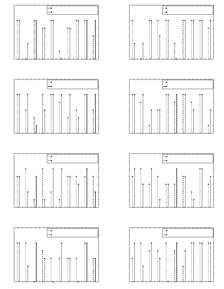
Using the data presented in the experimental
paradigm section, Fig.1 shows the number of pre-
pared contractions for each subject. The dashed (resp.
solid) line concerns the trial ”with” (resp. without”)
preparation warning. The results of Fig 1.a (resp. Fig
1.e) corresponds to male FDP (resp. female). Fig 1.b
(resp. Fig 1.f) represents the data of male FDS (resp.
female ). The results of Fig 1.c (resp. Fig 1.g) are
addressed to male FR (resp. female). Finally, the Fig
1.d (resp. Fig1.h) gives the number of preparation of
male CED s(resp. female).
Note that all volunteers prepare muscle activity
even when no preparation warning is given. How-
ever, the number of prepared contractions is not al-
ways the same. When comparing the muscles, we can
note that, in the case of males, the number of prepa-
ration is important for the flexor digitorum profundus
muscle (Fig 1.a) and for the flexor digitorum super-
ficialis muscle (Fig 1.b). In fact, 5 volunteers of 10
have a complete preparation (5 for both trials) during
contractions. The number of preparation decreases in
common extensor digitorum muscle to 3 preparations
(Fig 1.c). Hence, we can conclude that males have
an important number of preparation in flexor muscles
than in extensor muscles (Fig 1.a,b,c,d).
In the case of female volunteers, the number of prepa-
ration is more important in extensor muscles (Fig
1.g,h) than in flexor muscles (Fig 1.e,f). In fact,we
show one complete preparation in (Fig 1.f) but in (Fig
1.h), there is three complete preparations.
3.2 Analysis of the Number of Effective
Preparation
The objective of this section is to analyze the number
of effective preparation. To do it, this number is
used as input data. A simple statistical analysis using
the mean, the median and the standard deviation of
the number of preparation is carried and given in
Tab.1 in case of first radial muscle. We show that
male volunteers are characterized by an important
median and mean values of preparation number in
the case of ”without preparation”. But, the results are
opposite in case of female volunteers: the number
of preparation is slightly higher in case of ”with
icSPORTS 2015 - International Congress on Sport Sciences Research and Technology Support
42
1 2 3 4 5 6 7 8 9 10
0
1
2
3
4
5
6
7
Number of subjects
Number of preparation
With preparation warning
Without preparation warning
1 2 3 4 5 6 7 8 9 10
0
1
2
3
4
5
6
7
Number of subjects
Number of preparation
With preparation warning
Without preparation warning
(a) (b)
1 2 3 4 5 6 7 8 9 10
0
1
2
3
4
5
6
7
Number of subjects
Number of preparation
With preparation warning
Without preparation warning
1 2 3 4 5 6 7 8 9 10
0
1
2
3
4
5
6
7
Number of subjects
Number of preparation
With preparation warning
Without preparation warning
(c) (d)
1 2 3 4 5 6 7 8 9 10
0
1
2
3
4
5
6
7
Number of subjects
Number of preparation
With preparation warning
Without preparation warning
1 2 3 4 5 6 7 8 9 10
0
1
2
3
4
5
6
7
Number of subjects
Number of preparation
With preparation warning
Without preparation warning
(e) (f)
1 2 3 4 5 6 7 8 9 10
0
1
2
3
4
5
6
7
Number of subjects
Number of preparation
With preparation warning
Without preparation warning
1 2 3 4 5 6 7 8 9 10
0
1
2
3
4
5
6
7
Number of subjects
Number of preparation
With preparation warning
Without preparation warning
(g) (h)
Figure 1: Number of preparation for each subject. (a): male FDP, (b): male FDS, (c): male FR, (d): male CED, (e): female
FDP, (f): female FDS, (g): female FR, (h): female CED.
Development of EMG Indicators for Measuring and Analyzing Pre-motor Activity on Muscles
43
preparation warning” trial.
Table 1: Simple statistic values of number of preparation on
first radial muscle.
Mean Median Standard
deviation
Males With 3.8 4 1.55
Without 4 5 1.5
Females With 3.4 3 1.5
Without 3.1 3 1.96
Tab.2 represents the number of preparation per-
centage in both trials, for both genders and for each
muscle separately.
For male volunteers, the percentage of number of
preparation is important when no preparation warn-
ing is given. We observe this result in three muscles
(FDP, FDS, CED) except the first radial (FR) in case
of 5 preparations.
The sum of percentage of number of preparation is
equal to 80% for 3,4 and 5 contractions together.
This result is valid for all muscles except the com-
mon extensor digitorum muscle who had an impor-
tant percentage (100%) when no preparation warning
is given.
The results are opposite in the case of female vol-
unteers. We observed that the sum of percentage of
number of preparation are more important in case of
”with preparation warning” than in case of ”without
preparation warning” for 3,4 and 5 contractions to-
gether. We show this result in the three first mus-
cles: CED, FDS, FR. The first radial (FR) muscle had
90% (resp. 70%) preparations in case of ”with” (resp.
”without”) preparation, in FDS and CED .
However, for flexor digitorum profundus muscle, the
percentage is equal to 70% between both trials. Ac-
cording to Tab. 2, we noticed that males prepare more
than females in both trials.
4 THE EFFECT OF
PREPARATION’S DURATION
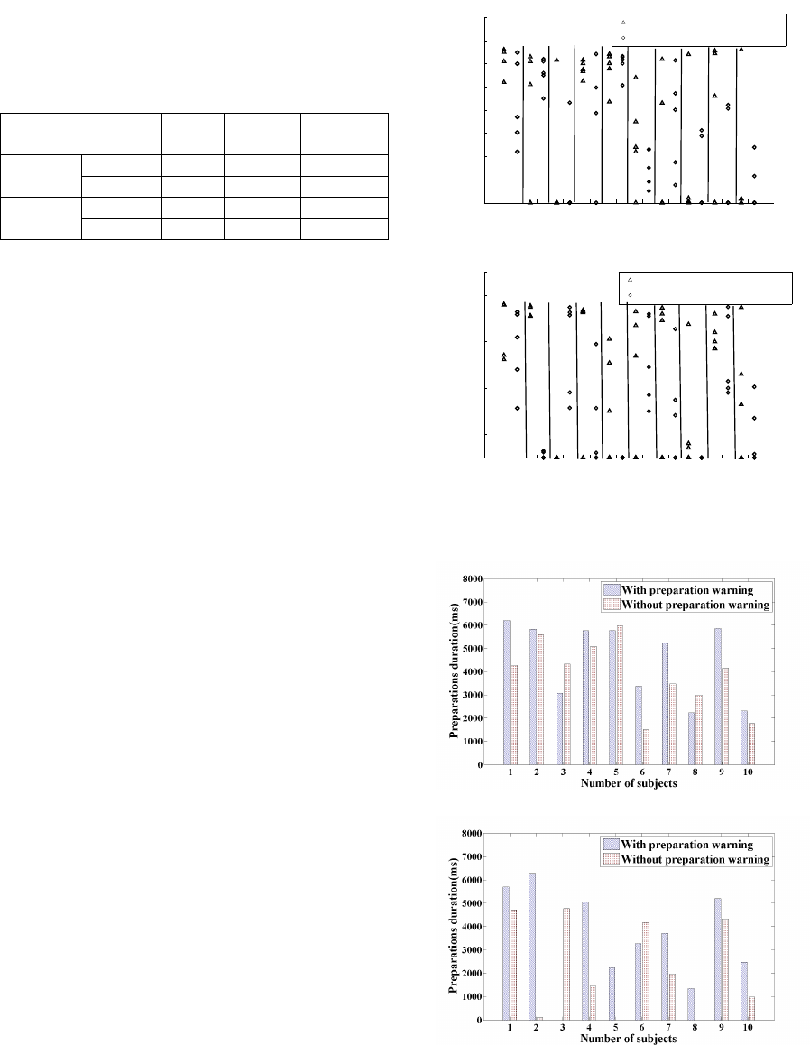
4.1 Duration by Subject
This section aims to observe the dispersion of prepa-
ration’s duration in the both trials.
Fig.2 shows the duration of preparation for each
male and female volunteers of first radial muscle. For
clarity reasons, symbols are discarded in the figure
from the central values. The triangle (resp. circle)
symbol represents ”With” (resp. ”Without”) prepara-
1 2 3 4 5 6 7 8 9 10
0
1000
2000
3000
4000
5000
6000
7000
8000
Number of subjects
Preparations duration (ms)
With preparation warning
Without preparation warning
(a)
1 2 3 4 5 6 7 8 9 10
0
1000
2000
3000
4000
5000
6000
7000
8000
Number of subjects
Preparations duration (ms)
With preparation warning
Without preparation warning
(b)
Figure 2: Preparation’s duration of first radial muscle.(a):
male volunteers,(b): female volunteers.
(a)
(b)
Figure 3: Mean preparation
´
s duration of first radial muscle.
(a): male volunteers, (b): female volunteers.
icSPORTS 2015 - International Congress on Sport Sciences Research and Technology Support
44

Table 2: Number of preparation percentage in both trials: with and without preparation warning.
Male volunteers Female volunteers
Number of preparations(%) 5 4 3 2 1 0 5 4 3 2 1 0
FDP With 50 20 10 0 10 10 20 50 0 10 20 0
Without 60 20 0 10 0 10 30 30 10 20 10 0
FDS With 70 10 0 10 0 10 20 30 40 10 0 0
Without 60 10 10 20 0 0 40 20 20 0 20 0
FR With 40 10 30 20 0 0 30 10 50 0 0 10
Without 70 0 10 10 10 0 40 0 30 10 0 20
CED With 40 30 10 0 20 0 50 30 10 10 0 0
Without 60 0 40 0 0 0 60 10 10 10 0 10
tion warning. These two genders have different val-
ues of preparation’s duration and don’t obey to any
obvious rules. Note that the range of all durations is
between 0 and 6600 milliseconds (we recall that the
duration of theoretical preparation is 6600 millisec-
onds).
Fig.3 illustrates the mean preparation’s duration
for each trial and for every volunteer. Fig 3.a (resp.
Fig 3.b) represents males (resp. female) volunteers.
The mean preparation’s duration is higher in case of
”with preparation warning” trial for 7 males and 9 fe-
males. Hence, we can conclude that volunteers pre-
pare longer their contraction when a warning signal is
given.
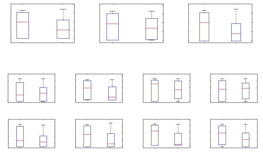
4.2 Boxplots of Preparation’s Duration
In statistic analysis, the boxplots is a useful tool for
studying large sets of data. It can provide informa-
tion about data range, median, normality and skew of
distribution. In this case, we deal with the distribu-
tion of preparation’s duration for the two trials for all
muscles.
In Fig. 4, we draw the preparation’s duration boxplots
for both gender when all muscles data are combined
together. We can see that the dispersion is large when
a preparation warning is given. For example, in Fig
4.a, the median duration value (line into rectangle) of
pre-motor activity is important in ”with preparation
warning” (3.83 s) than ”without preparation warning”
mode (1.987 s).
Fig.5 represents the preparation’s duration for each
muscle for male and female volunteers separately.
The results shows that the median duration value is
higher in ”with preparation warning” trial than in
”without preparation warning” trial except the Fig5.a
and Fig5.d who show that the median value is slightly
higher when no preparation warning is given.
5 STATISTIC ANALYSIS: ONE
WAY ANOVA TEST
In statistics, ANalysis Of VAriance (ANOVA)(Nuzzo,
2014) is a collection of statistical models used in or-
der to analyze the differences between group means
and their associated procedures (such as ”variation”
among and between groups). Anova test was used
to compare the preparation’s duration and try to sepa-
rate the two trials with significance level equal to 5%.
Tab.3 gives the results of discrimination between
two trials. The symbol (6= ) (resp. (=)) means there is
a (resp. no) significant difference between two trials.
Table 3: Difference inter-muscular between with and with-
out preparation warning.
Males Females Both gender
All muscles = 6= 6=
FDP = = =
FDS = 6= 6=
FR = 6= 6=
EDC = 6= =
5.1 Difference between ”With
preparation warning” and ”Without
preparation warning” on Muscles
The results of Tab.3 show that there is a signifi-
cant difference between with and without preparation
warning for both genders and for all muscles con-
sidered together(T=14.48,p-value=0.0001). So, the
preparation warning has an influence on the pre-motor
activity.
Development of EMG Indicators for Measuring and Analyzing Pre-motor Activity on Muscles
45
With Without
0
2000
4000
6000
8000
Preparations duration (ms)
With Without
0
2000
4000
6000
8000
Preparations duration (ms)
With Without
0
2000
4000
6000
8000
Preparations duration (ms)
(a) (b) (c)
Figure 4: Boxplots of preparation’s duration for forearm muscles. (a):both gender, (b):male volunteers, (c):female volunteers.
With Without
0
2000
4000
6000
8000
Preparation’s duration (ms)
With Without
0
2000
4000
6000
8000
Preparation’s duration (ms)
With Without
0
2000
4000
6000
8000
Preparation’s duration (ms)
With Without
0
2000
4000
6000
8000
Preparation’s duration (ms)
(a) (b) (c) (d)
With Without
0
1 000
2 000
3 000
4 000
5 000
6 000
7 000
8 000
Preparation’s duration (ms)
With Without
0
2000
4000
6000
8000
Preparation’s duration (ms)
With Without
0
2000
4000
6000
8000
Preparation’s duration (ms)
With Without
0
2000
4000
6000
8000
Preparation’s duration (ms)
(e) (f) (g) (h)
Figure 5: Boxplots of preparation
´
s duration for forearm muscles. (a): male FDP, (b): male FDS, (c): male FR, (d): male EDC
(e): female FDM, (f): female FDS, (g): female FR, (h): female EDC.
5.2 Gender Difference in Pre-motor
Activity
Tab.3 shows that preparation’s duration differs by
gender. In fact, there is no difference between with-
out and with preparation warning for males (T=1.71,
p-value=0.1922) but there is a significant difference
in females (T=16.32, p-value=0.00005) for all mus-
cles. These results confirm the brain behavior. In fact,
in some previous studies such as (Ingalhalikar et al.,
2014), it was shown that the brain behavior is differ-
ent for each gender: the females outperform males
on attention, word and face memory, and social cog-
nition tests and males perform better on spatial pro-
cessing and motor and sensorimotor speed. In this
case and specially in ”with preparation warning” ,vol-
unteers must be attentional and concentrate. Female
were able to do it. However, male volunteers weren’t
sensitive to preparation warning.
5.3 Inter-muscular Difference in
Pre-motor Activity
When we separate the muscles, the results of Tab.3
show that there is no difference between with and
without preparation warning in pre-motor activity for
males (p-value ≥ 0.134). For females, we found
a significant difference in Flexor Digitorum Super-
ficialis, First Radial , Extensor Digitorum Commu-
nis muscles (T ≥ 4.72, p-value ≤ 0.032) but no dif-
ference in Flexor Digitorum Profundus (T=2.29, p-
value=0.1302).
The muscles are classified into two families: the
superficial muscles and deep muscles. The superfi-
cial (resp. deep) muscles are Flexor Digitorum Su-
perficialis, First Radial and Extensor Digitorum Com-
munis (resp. Flexor Digitorum Profundus). Due to
the anatomical and biomechanical differentiation of
the superficial and deep muscle fibers, a difference
in fiber type distribution can be hypothesized: it is
assumed that the deep muscle fiber has a higher por-
tion of type I fibers compared to the superficial mus-
cle fiber (MacDonald et al., 2006). Fibers of type I are
slow twitch fibers, which are fatigue resistant and ide-
ally suited to provide low load tonic activity. Type II
fibers, are fast twitch fibers, are less fatigue resistant,
but able to produce a higher load activity(Henneman
et al., 1965).
During the pre-motor activity, the preparation was
done only for superficial muscles.
icSPORTS 2015 - International Congress on Sport Sciences Research and Technology Support
46
6 CONCLUSION
In this work, we found two indicators that character-
ize the pre-motor activity. The first one is the number
of preparation. It was shown to be important even
when no preparation warning is given.
The second indicator is preparation’s duration. It was
shown a significant difference between ”with” and
”without” preparation modes for female in superficial
muscles of the forearm. However, no difference of
behavior are observed between the two modes for
male.
Studying the motor behavior during the transition
between the pre-motor activity and the effective
motor activity and studying the brain activity using
fMRI will be the topic of further research.
REFERENCES
Basmajian, J. V. (1979). Biofeedback: Principles and prac-
tice for clinicians. Williams & Wilkins.
Bøg, M. F., Erkocevic, E., Niemeier, M. J., Mathiesen, J. R.,
Smidstrup, A., and Kamavuako, E. N. (2011). In-
vestigation of the linear relationship between grasp-
ing force and features of intramuscular emg. In 15th
Nordic-Baltic Conference on Biomedical Engineering
and Medical Physics (NBC 2011), pages 121–124.
Springer.
Cacioppo, J. T., Tassinary, L. G., and Berntson, G. (2007).
Handbook of psychophysiology. Cambridge Univer-
sity Press.
Corbin, C. B. (1967). Effects of mental practice on skill de-
velopment after controlled practice. Research Quar-
terly. American Association for Health, Physical Edu-
cation and Recreation, 38(4):534–538.
Gordon, R., Graham, C., and Joseph, H. (2004). Research
Methods in Biomechanics. Human Kinetics.
Henneman, E., Olson, and B., C. (1965). Relations between
structure and function in the design of skeletal mus-
cles. Journal of Neurophysiology, 28(3):581–598.
Ingalhalikar, M., Smith, A., Parker, D., et al. (2014). Sex
differences in the structural connectome of the human
brain. Proceedings of the National Academy of Sci-
ences, 111(2):823–828.
MacDonald, D. A., Moseley, Lorimer, G., H., and W., P.
(2006). The lumbar multifidus: does the evidence sup-
port clinical beliefs. Manual therapy, 11(4):254–263.
Nuzzo, R. (2014). Scientific method: Statistical errors. nat,
506:150–152.
Richardson, A. (1967). Mental practice: a review and dis-
cussion part i. Research Quarterly. American Associ-
ation for Health, Physical Education and Recreation,
38(1):95–107.
Shelton, T. O. and Mahoney, M. J. (1978). The content
and effect of psyching-up strategies in weight lifters.
Cognitive Therapy and Research, 2(3):275–284.
Weinberg, R. S. (1981). The relationship between mental
preparation strategies and motor performance: A re-
view and critique. Quest, 33(2):195–213.
Development of EMG Indicators for Measuring and Analyzing Pre-motor Activity on Muscles
47
