Extraction of Pectin from Durian Rind and Its Minimum Inhibitory
Concentration towards Staphylococcus Aureus and Escherichia Coli
Mufti Ghaffar
1
, Harsi Dewantari Kusumaningrum
2
and Nugraha Edhi Suyatma
2
1
Study Program of Food Science, Graduate School, IPB University, Bogor 16680, Indonesia
2
Department of Food Science and Technology, IPB University, Bogor 16680, Indonesia
Keywords: Durian Rind, Macrodilution, MIC, Pectin Isolate.
Abstract: Durian is a big tropical fruit grown in Indonesia that leaves many waste from its rind. Recently, some reports
shows that durian rind can be extracted for its pectin that showing a potential antimicrobial activity toward
pathogen. The aim of this study was to characterized durian pectin isolate from two different extraction
methods and to determine the minimum inhibitory concentration (MIC) of durian pectin isolate against
pathogenic bacteria. Inhibitory activity of durian pectin isolate against two bacterial strain: Staphylococcus
aureus and Escherichia coli, was determined by using macrodilution methods, and amoxicilline used as
positive control. Results showed that water content of the two extraction methods resulted in around 96%.
Yield of durian pectin isolate in methods A (5,45 ± 0,10 %) more than extraction B (3,46 ± 0,06 %). MIC of
isolates durian pectin in mueller hinton broth (MHB) medium against E. coli was 500 mg/ml and S. aureus
was 110 mg/ml, this concentration showed no sign of bacterial growth, this condition was same as positive
control.
1 INTRODUCTION
Indonesia is tropical country that has many fruit can
grow, one of that fruit is Durian (Figure 1a). Durian
is a popular fruit in Indonesia, a tropical, seasonal
climateric fruit belonging to the Bombacaceae family.
Durian fruit is quite large, round or oval, green
(brownish when ripe), has a thick outer shell, hard,
and covered with many thorns shaped like a pyramid.
Durian fruit has a short shelf life, it is known to be
damaged 36 to 72 hours from the time the durian fruit
falls (Manoharan, 2013). Durian grows well in 75-
80% humidity conditions with rainfall between 1600
and 4000 ml a year, and with an average temperature
of 24-30
o
C. Requires a tropical climate to grow, and
not grow well in areas over 3000 feet (Ashraf et al.,
2011).
It is customary for people in several regions in
Indonesia to use durian rind as a drinking container
after eating durian fruit. It is said that by drinking
water from durian rind can eliminate the pungent odor
of durian fruit, and can reduce durian motion sickness
due to consumption of durian in large quantities.
Based on this, there is a possibility that the durian rind
contains active components, but has not been much
studied. It is reported in some literature that durian
rind has a number of therapeutic benefits such as: has
anti-diabetic properties, anti-hyperlipidemic effect,
anti-proliferative activity, and antimicrobial activity.
Pectin can be extracted from durian rind which has
antimicrobial activity. Pectin from durian rind has the
ability to inhibit the growth of Vibrio harveyi 1526
(MIC = 6.3 and 12.5 mg / mL) in black tiger / tiger
shrimp (Pholdaeng and Pongsamart, 2010). Durian
rind pectin has anti microbial activity against E. coli, S.
aureus (Lipipun et al., 2002). Films made from durian
rind pectin also have antibacterial properties when
tested on several bacteria (Ho and Bhat, 2015).
Utilization of durian rind has not been done much
by households, or industries that process durian fruit.
Pectin can be extracted from durian rind which has
antimicrobial properties that have the potential to be
developed. This study aims to characterized durian
pectin isolate and to determine the minimum
inhibitory concentration of durian pectin isolate
against pathogenic bacteria E. coli and S. aureus.
2 MATERIALS AND METHODS
2.1 Materials
Durian was obtained from the Warso Farm durian
72
Ghaffar, M., Kusumaningrum, H. and Suyatma, N.
Extraction of Pectin from Durian Rind and Its Minimum Inhibitory Concentration towards Staphylococcus Aureus and Escherichia Coli.
DOI: 10.5220/0009978600002833
In Proceedings of the 2nd SEAFAST International Seminar (2nd SIS 2019) - Facing Future Challenges: Sustainable Food Safety, Quality and Nutrition, pages 72-76
ISBN: 978-989-758-466-4
Copyright
c
2022 by SCITEPRESS – Science and Technology Publications, Lda. All rights reserved
farm, Bogor, Indonesia. Staphylococcus aureus
ATCC 25923 and Escherichia coli ATCC 25922 use
as test bacteria were obtained from the food
microbiology laboratory of Food Science and
Technology Department IPB University. The initial
treatment of raw materials begins with durian rind cut
and separated mesocarp (the inner part of durian rind)
and exocarp (the outer part of durian rind). Mesocarp
was cut to a size of ± 0.5 cm to maximize the process
of drying and crushing the material. The durian peel
was placed into aluminum foil and dried in an oven at
60
o
C for 48 hours, then crushed to powder and
wrapped in adhesive plastic or stored in a desiccator.
2.2 Methods
2.2.1 Durian Pectin Extraction
The extraction method in this study uses the
extraction method from Amaliah (2014) and
Hokputsa et al. (2004) and compared the
characteristics of durian rind pectin isolates obtained.
The following was the Amaliah extract method
(2014) with a modification (extraction A). The
process of extracting pectin from durian rind is
carried out through several stages, heating, filtering,
concentrating, extracting. Durian rind powder was
mixed with distilled water (durian rind powder:
aquades = 1: 25 w / v) and stirred until it becomes an
acidic solution with a pH of 2 by adding 2 N HCl acid
solution and extracted at 85
o
C for 2 hours. Then
filtered using a cloth, then added an acid-ethanol
solution (4% HCl in 95% ethanol) with a ratio of 1: 1
v / v and incubated at room temperature for 1 hour.
To separate the gel (durian pectin isolate) filtering is
done in vacuum conditions. The resulting gel is rinsed
twice using 95% ethanol (1: 1, v / v) and shaken for
15 minutes (Amaliyah, 2014).
The following was a method of extraction
Hokputsa et al. (2004) with the modification
(extraction B). Durian skin powder was mixed with
distilled water (durian skin powder: aquades = 1: 25
b / v) and stirred to form an acidic solution with a pH
of 4.5 with citric acid solution added and extracted at
a temperature of 90-100
o
C for 20 minutes. Then
filtered using a cloth, then added an acid-ethanol
solution (4% HCl in 95% ethanol) with a ratio of 1: 1
v / v and incubated at room temperature for 1 hour.
To separate the gel / durian pectin solate filtering
done by vacuum conditions. The resulting gel is
rinsed twice using 95% ethanol (1: 1, v / v) and
shaken for 15 minutes. The resulting sludge was a
durian pectin isolate (DPI). The DPIs obtained were
then characterized in the form of measurements of
water content, yield, and pH values.
2.2.2 Measurement Antibacterial Activity
from Durian Pectin Isolate
Microorganism that used in this macrodilution
method are common contaminating microorganism in
food products, namely: Eschericia coli and
Staphylococcus aureus. Macrodilution is one of the
basic antimicrobial testing methods. Before the
macrodilution was carried out the sterilization
process was done on samples of DPI. The procedure
begins by testing an antimicrobial agent DPI with a
concentration of 1) 100%, 50%, 25%, 12.5%, 6.25%,
3.125% (E. coli) and 2) 100%, 25%, 6,25%, 1,56%,
0,39%, 0,09 % (S. aureus) in 2 mL (E. coli) and 3 mL
(S. aureus) microbial growth medium (Mueller
Hinton Broth) in a tube. Then each tube was
inoculated with a bacterial inoculum prepared in the
same medium. The size of the inoculum microbe is
around 5 x 10
5
CFU / mL. After mixing well, each
tube was incubated at 30
o
C for 20 hours. Then each
tube was inoculated on the Mueller Hinton Agar
medium by scratching, because DPI covers the
detection of microbial growth with its color. This
procedure was repeated three times (Balouiri et al.,
2016).
3 RESULTS AND DISCUSSIONS
3.1 Durian Pectin Isolate
Characterization
To get durian pectin isolate, the initial preparation
process of durian rind raw material was done to
separate the mesocarp (Figure 1b) and exocarp. From
this part (mesocarp) had yield approximately 29.63 ±
0.02% from all part of durian rind. Mesocarp is a raw
material that used to obtain durian rind powder. And
the yield of durian rind powder was 10,44 ± 0.06
(Table 1).
Table 1: Yield and water content of mesocarp and durian
powder.
Sample Yield (%)
Water
content (%)
Mesocarp 29,63 ± 0.02 -
Durian rind powder 10,44 ± 0.06 1,01 ± 0,17
Extraction of Pectin from Durian Rind and Its Minimum Inhibitory Concentration towards Staphylococcus Aureus and Escherichia Coli
73
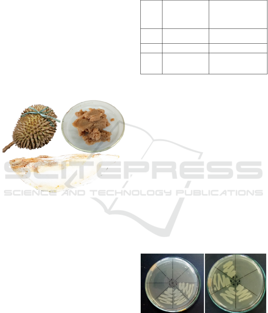
Characterization of durian pectin isolate (Figure
1c) was performed, giving values of water content,
yield and pH value (Table 2). Water content of both
methods resulted about 96%% which indicated that
durian pectin isolate was composed of large amounts
of water. Extraction method A (pH: 2.00,
temperature: 85
o
C, t: 2 hours) has bigger yield (5,45
± 0,10) than extraction B (pH: 4.5 and temperature:
90-100
o
C, t : 20 minutes that had yield 3,46 ± 0,06,
which indicates extraction A had more yield than
extraction B. The more extraction yield are related to
differences of pH. The lower the pH, the more yield
of pectin will be obtained. The yield of both methods
is in the range of the previous report's yield, which
ranges from 1.04% - 10% (Arlofa et al., 2015;
Pholdaeng and Pongsamart, 2010; Wai et al., 2009).
Figure 1: a) Durian fruit, b) Mesocarp of durian fruit, and
c) Durian pectin isolate.
Lower pH value result in more yield of pectin can
be extracted (Ardiansyah et al., 2014). The principle
of extraction of pectin is the overhaul of protopectin
which does not dissolve into pectin which can
dissolve with acid solvents. In the extraction process
with a high level of acidity will increase the
hydrolysis of protopectin from durian rind tissue to be
dissolved in water faster, so that the yield of pectin is
higher. Protopectin does not dissolve easily because
it is in the form of calcium and magnesium salts.
Hydrolysis of protopectin with acids causes hydrogen
ions to replace calcium ions and magnesium ions in
the protopectin molecule. This is due to a pH of 1.5
which is more acidic and has more hydrogen ions so
that the possibility of substituted calcium and
magnesium is greater (Prasetyowati et al., 2009).
Table 2: Yield, pH, and water content from extraction
method A and extraction method B.
Para-
meter
Extraction A
(acid: HCl 2 N, pH:
2, Temperature:
85
o
C, time: 2 hour)
Ekstraction B
(acid: citric acid, pH: 4.5,
Temperature: 90-100
o
C,
time: 20 minutes)
Yield
(%)
5,45 ± 0,10 3,46 ± 0,06
pH 2,03 ± 0,10 3,75 ± 0,09
Water
content
(%)
96,26 ± 0,1 96,4 ± 0,04
3.2 Antibacterial Activity of Durian
Pectin Isolate
Durian pectin isolates (DPI) obtained through the
extraction process were tested for their antibacterial
activity. The test results showed that there were two
concentrations of DPI and positive control that could
inhibit the E.coli bacteria as indicated by the clear
growth media (Figure 1). Both concentrations are
100% and 50% for E. coli and concentration 100%
and 25 % for S.aureus. From that figure we can get
the value of the minimum inhibitory concentration
(MIC) of each bacteria (Table 3). Based on the results
it was found that the MIC value of DPI in E. coli was
500 mg/ml and S. aureus was 110 mg/ml.
The difference in antibacterial activity that occurs
in Gram-positive (S. aureus) and Gram-negative (E.
coli) bacteria is probably caused by differences in the
composition and structure of cell walls in the two
types of bacteria. The structure of the cell wall of
Gram-positive bacteria is simpler, that is single layer
with a low lipid content (1-4%) making it easier for
bioactive materials to enter the cell. The structure of
the cell wall of Gram negative bacteria is more
Figure 2: a) Growth Escherichia coli inhibited by durian
pectin isolate in MHA medium (1: 100%, 2: 50%, 3: 25%,
4: 12.5%, 5: 6.125 %, 6: clear media, 7: amoxicilin as
positive control), b) Growth Staphylococcus aureus
inhibited by durian pectin isolate in MHA medium
(1: 100%, 2: 25%, 3: 6,25%, 4: 1,56%, 5: 0,39 %.
a)
b)
c)
a
b
2nd SIS 2019 - SEAFAST International Seminar
74
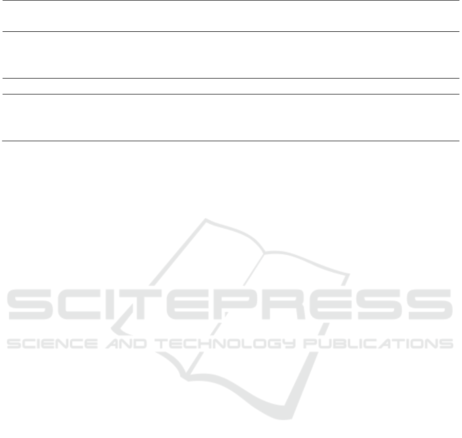
Table 3: Microbial growth and minimum inhibitory concentration of durian pectin isolate against E. coli dan S. aureus.
Bacteria Antibacterial concentration (%) MIC (%)
100 50 25 12,5 6,25 positive control
E. coli 1 - - ++ ++ ++ - 50
2 - - ++ ++ ++ - 50
3 - - ++ ++ ++ - 50
100 25 6,25 1,56 0,39 positive control
S. aureus 1 - + ++ ++ ++ - 25
2 - - ++ ++ ++ - 25
3 - - ++ ++ ++ - 25
- : no growth or no colony
+ : there is growth or there is colony
++ : many growth or many colony
bioactive materials to enter the cell. The structure of
the cell wall of Gram negative bacteria is more
complex, three layers, namely the outer layer of
lipoprotein, the middle layer of lipopolysaccharide
which acts as a barrier to the entry of antibacterial
bioactive material, and the inner layer is in the form
of peptidoglycan with high lipid content (11-12%)
(Magdalena and Kusnadi, 2015).
4 CONCLUSIONS
Durian pectin isolate of both extraction methods
resulted in water content aproximately 96 %.
Extraction method A (pH: 2.00, temperature: 85
o
C, t:
2 hours) had bigger yield (5,45 ± 0,10) than extraction
B (pH: 4.5 and temperature: 90-100
o
C, t: 20 minutes)
that had yield 3,46 ± 0,06 %. Minimum inhibitory
concentration of durian pectin isolate against
pathogenic bacteria E. coli was 500 mg/ml and S.
aureus was 110 mg/ml.
ACKNOWLEDGEMENT
Thanks to Beasiswa Unggulan Kemendikbud
Republik Indonesia that sponsored author study in
IPB University.
REFERENCES
Amaliyah, D.M., 2014. Pemanfaatan Limbah Kulit Durian
(Durio zibethinus) dan Kulit Cempedak (Artocarpus
integer) sebagai edible film. J. Ris. Ind. Has. Hutan 6,
27–34.
Ardiansyah, G., Hamzah, F., Efendi, R., 2014. Variasi
Tingkat Keasaman Dalam Ekstraksi Pektin Kulit Buah
Durian. JOM FAPERTA 1.
Arlofa, N., Safuari, A., Gunawan, I.G., 2015. Pektin Kulit
Durian Sebagai Bahan Baku Biosorben Logam Berat
Timbal (Pb). J. Chemtech 1, 6–11.
Ashraf, M.A., Maah, M.J., Yusoff, I., 2011. Estimation of
Antioxidant Phytochemicals in Four Different Varieties
of Durian. Int. Conf. Biosci. Biochem. Bioinforma. 5,
131–135.
Balouiri, M., Sadiki, M., Ibnsouda, S.K., 2016. Methods for
in vitro evaluating antimicrobial activity: A review. J.
Pharm. Anal. 6, 71–79. https://doi.org/10.1016/j.jpha.
2015.11.005
Ho, L.H., Bhat, R., 2015. Exploring the potential
nutraceutical values of durian (Durio zibethinus L.) -
An exotic tropical fruit. Food Chem. 168, 80–89.
https://doi.org/10.1016/j.foodchem.2014.07.020
Hokputsa, S., Gerddit, W., Pongsamart, S., Inngjerdingen,
K., Heinze, T., Koschella, A., Harding, S.E., Paulsen,
B.S., 2004. Water-soluble polysaccharides with
pharmaceutical importance from Durian rinds (Durio
zibethinus Murr.): isolation, fractionation, characterisa-
tion and bioactivity. Carbohydr. Polym. 56, 471–481.
Lipipun, V., Nantawanit, N., Pongsamart, S., 2002.
Antimicrobial activity ( in vitro ) of polysaccharide gel
from durian fruit-hulls. Songklanakarin J. Sci. Technol.
24, 31–38.
Magdalena, N.V., Kusnadi, J., 2015. Antibakteri dari
ekstrak kasar daun gambir (Uncaria gambir var
Cubadak) metode microwave-assisted extraction
terhadap bakteri patogen. J. Pangan dan Agroindustri 3,
124–135.
Manoharan, S., 2013. Synergistic Activity of Chloroform
Extract of Durio zibethinus Wood Bark With Penicillin
G Against Staphylococcus aureus . Int J Biol Med Res.
3, 3025–3027.
Pholdaeng, K., Pongsamart, S., 2010. Studies on the
immunomodulatory effect of polysaccharide gel
extracted from Durio zibethinus in Penaeus monodon
shrimp against Vibrio harveyi and WSSV. Fish
Extraction of Pectin from Durian Rind and Its Minimum Inhibitory Concentration towards Staphylococcus Aureus and Escherichia Coli
75
Shellfish Immunol. 28, 555–561. https://doi.org/
10.1016/j.fsi.2009.12.009.
Prasetyowati, Sari, K.P., Pesantri, H., 2009. Ekstraksi
Pektin Dari Kulit Mangga. 42 J. Tek. Kim. 16, 42–49.
Wai, W.W., Alkarkhi, A.F.M., Easa, A.M., 2009.
Optimization of pectin extraction from durian rind
(durio zibethinus) using response surface methodology.
J. Food Sci. 74, 637–641. https://doi.org/10.1111/
j.1750-3841.2009.01331.x.
2nd SIS 2019 - SEAFAST International Seminar
76
