
A Case Report: Psoriasis-mimicking Lesions of Secondary Syphilis
in HIV Positive Patient
Vika Fintaru
1*
, Sunardi Radiono
2
, Satiti Retno Pudjiati
2
1
Resident of Dermatology & Venereology Department, Faculty of Medicine, Public Health & Nursing, Universitas Gadjah
Mada/Dr. Sardjito General Hospital, Yogyakarta
2
Dermatology & Venereology Department, Faculty of Medicine, Public Health & Nursing, Universitas Gadjah Mada/Dr.
Sardjito General Hospital, Yogyakarta
Keywords: Psoriasis, secondary syphilis, HIV
Abstract: Syphilis is a sexually transmitted disease that is considered to be "the great imitator" because of the
clinical appearance that resembles various types of skin diseases. The coincidence between syphilis and
Human Immunodeficiency Virus(HIV) often occurs, and the incidence increases, especially in the male
population who have sex with men. Uncommon syphilis clinical manifestations are common in HIV
patients. This paper reports a 28-year-old man who was infected with HIV, came to Dr. Sardjito
General Hospital's Dermatovenereology polyclinic, with a complaint of scaly red plaques on the arms,
legs, hands, feet, and scrotum and accompanied by nail abnormalities that mimicking skin and nail
abnormalities in psoriasis. Histopathology examination appropriate with secondary syphilis with
psoriasiform acanthosis, neither hypergranulosis nor Monroe abscess, and many lymphocytes and
plasma cells infiltration in the upper dermis, perivascular, and periadnexa. The serological test showed
a positive result of Treponema Pallidum Haemagglutination (TPHA) and Venereal Disease Research
Laboratory (VDRL) 1/32. The patient was treated with injections of benzathine penicillin G 2.4 million
units single dose. Skin and nail lesions improvement occurred three months after therapy. Variable
clinical presentations of secondary syphilis in HIV disease may lead to wrong diagnosis and improper
treatment. The biopsy can be used to make a diagnosis of atypical syphilis lesions. Syphilis patients
with HIV infectionmore likely to experience serological declinefailure, recurrent infections, and slower
treatment response than patients who are not infected with HIV.
1 INTRODUCTION
Syphilis is a chronic sexually transmitted disease
caused by Treponema pallidum subspecies pallidum.
Syphilis can affect almost all of the internal organs,
including cardiovascular and nervous systems.
Transmission most often occurs through direct
contact with lesions in mucosa or skin during sexual
intercourse through vagina, anus, or genital. The
clinical manifestations of syphilis may resemble a
variety of diseases so often called "the great
imitator". (Zetola et al., 2002)
Syphilis prevalence in HIV patients is higher
than Human Immunodeficiency Virus (HIV)
negative patients. The coincidencebetween syphilis
and HIV often occurs because both are transmitted
primarily through sexual intercourse. (Karp et al.,
2009) Syphilis patients with ulcer lesions are at
higher risk of HIV infection than other syphilis
lesions. The high rates of coincidence of syphilis and
HIV make all patients with syphilis should be
offered HIV testing, and all HIV patients must
regularly undergo syphilis examinations, especially
for high-risk populations.
(Pastuszczak et al.,2011 )
According to World Health Organization
(WHO) data, there are around 900,000 new cases of
syphilis every year so the clinician must be aware of
the disease (Karp et al., 2009). Since 2014-2018,
Dr. Sardjito General Hospital has found 114 cases
of syphilis with 53 cases of syphilis coincidence
with HIV. This paper reports a case of psoriasis-
mimicking lesions of secondary syphilis in
homosexual patients with HIV.
Fintaru, V., Radiono, S. and Pudjiati, S.
A Case Report: Psoriasis-mimicking Lesions of Secondary Syphilis in HIV Positive Patient.
DOI: 10.5220/0009988103330337
In Proceedings of the 2nd International Conference on Tropical Medicine and Infectious Disease (ICTROMI 2019), pages 333-337
ISBN: 978-989-758-469-5
Copyright
c
2020 by SCITEPRESS – Science and Technology Publications, Lda. All rights reserved
333
2 CASE
A 28-year-old man came to the Dermatology and
Venereology Polyclinic of Dr. Sardjito General
Hospital Yogyakarta on May 2
nd
, 2018
complainedred and scaly plaques, felt itchy on the
palms and feet soles since nine months ago. Plaques
also appeared on the back of the hands, instep,
forearms, legs, and scrotum. Two months before,the
patient said that there had been a painlessulcer and
untreated, but improved by itself. The skin around
the nails began reddishly, and there were pitting
nails. The patient did not have a previous similar
skin disease, and atopy history was denied. The
patient does not have serious illnesses or take
routine medication. The same complaint from the
family is also denied. The patient was diagnosed
with HIV since December 2015 and started taking
antiretroviral therapy(ARV) since January 2018
using fixed drug combination(FDC) tenofovir,
hiviral, and efavirenz with 162/ml CD4 count. The
patient had unsafe sexual intercourse with ten men,
insertive, and receptively.
On the physical examination, it was found
that the patient's condition was moderate, compos
mentis, all vital sign within standard limit and no
lymph nodes enlargement. Dermatological and
venereological status of palmar manus and plantar
pedis appeared diffuse erythematous plaques with
thick scales, on the arms, lower limbs, dorsum
manus, and dorsum pedis appeared erythematous
plaques with thick scales, multiple and scattered, on
the scrotum appearedscaleswith excoriation, and on
nails appeared periungual erythema, discoloration,
onychodystrophy and pitting nails were obtained
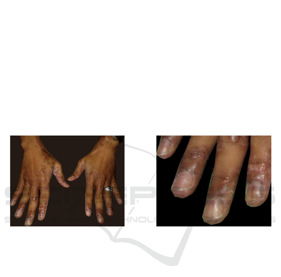
(Figure 1 and 2).
Figure 1: Dorsum manus: erythematous plaques with a
thick scaleon it, multiple, discrete
Figure 2: Dorsum manus (high magnification): periungual
erythema nails, discoloration, onychodystrophy and pitting
nails
Syphilis serological examination was performed
with a positive result of Haemagglutination
Treponema Pallidum (TPHA) and Venereal Disease
Research Laboratory (VDRL) 1/32 and patient was
HIV patient thus confirming the diagnosis of
secondary syphilis and coincidence with HIV. The
result of routine blood, liver function, kidney
function, and an electrolyte within reasonable limit.
Histopathological features of skin biopsy in the right
arm and right leg with hematoxylin-eosin (HE)
staining showed basket weave's type orthokeratosis,
psoriasiform acanthosis, spongiosis, minimal basal
cell vacuolar degeneration, and none of
hypergranulosis. For the dermis, patchy type of
inflammatory cell infiltration was obtained, mainly
lymphocytes and plasma cells in the upper dermis,
perivascular, and periadnexa (Figure 3A and 3B).
Histopathological results were in accordance with
secondary syphilis.
ICTROMI 2019 - The 2nd International Conference on Tropical Medicine and Infectious Disease
334
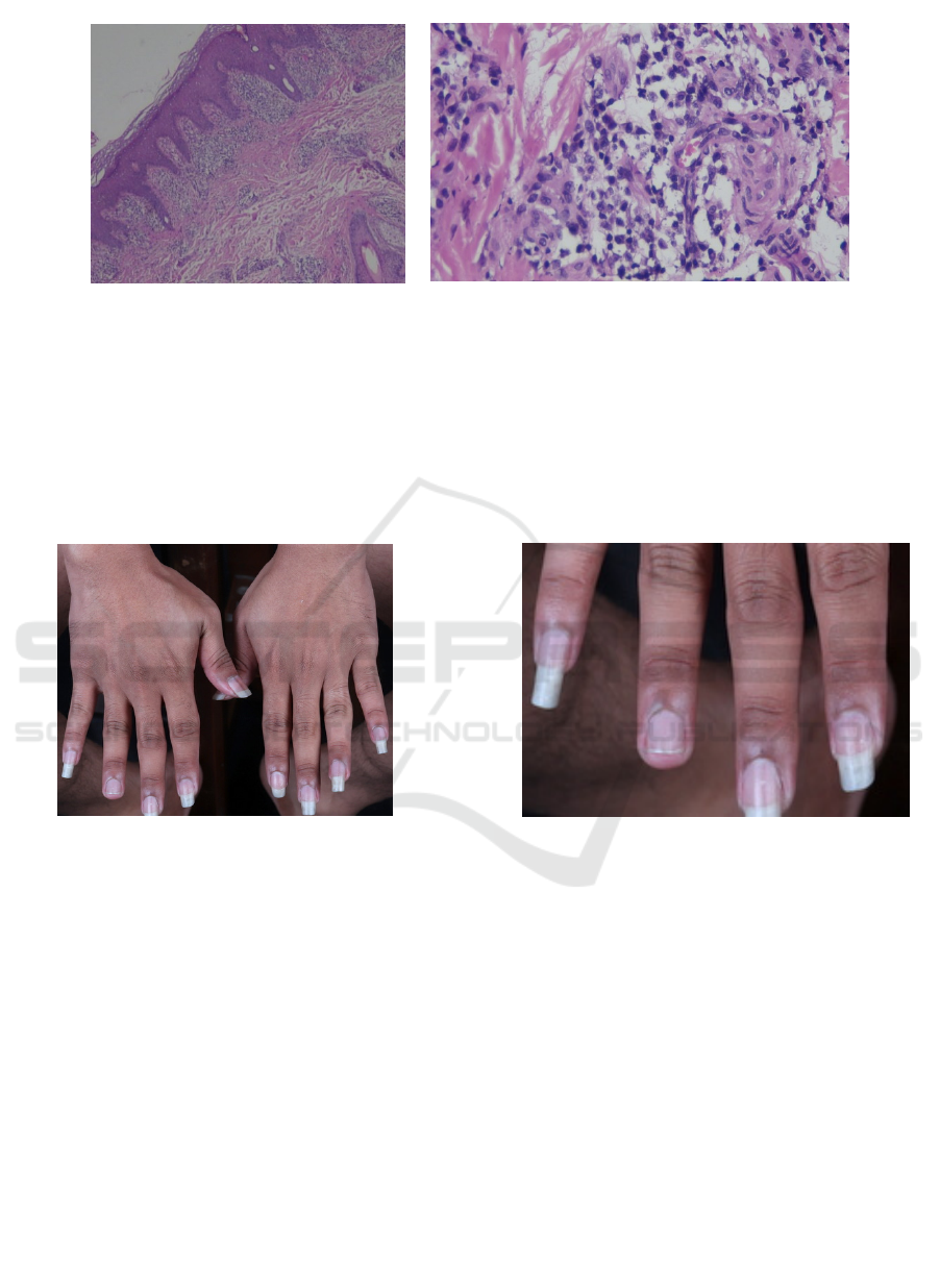
3A
3B
Figure 3: Histopathologic figures of secondary syphilis. A. basket weave's type orthokeratosis, psoriasiform acanthosis,
spongiosis, minimal basal cell vacuolar degeneration, and none hypergranulosis B. Highest magnification: many plasma
cells in the upper dermis
Diagnosis of psoriasiform type secondary
syphilis confirmed based on patient medical history,
physical examination, and histopathological
examination. Treatment was started with a single
dose of 2.4 million unit benzathine penicillin G. One
month after therapy, skin lesions and nails also
VDRL serological examination did not decline. Skin
and nail lesions improvement occurred in the 3
rd
month after treatment, but VDRL was still 1/32 until
six months after treatment. In the 9
th
month after
treatment, there was a decrease in VDRL into 1/16.
Figure 4: after being given standard therapy, skin lesions
improved, subungual erythema, discoloration, an
d
onychodystrophy improved, there were still a few pitting
nails in digit II and III manus dextra
Figure 5: after being given standard therapy, skin lesions
improved, subungual erythema, discoloration, an
d
onychodystrophy improved, there were still a few pitting
nails in digit II and III manus dextra
3 DISCUSSION
Syphilis is a chronic infectious disease caused by
spirochete Treponema pallidum, a spiral-shaped
bacterium with two until three flagella at each end.
Syphilis spreads through direct contact with the
lesion, although a small part of infections spread by
blood transfer, for example, during blood
transfusions or sharing needles toinject the drug. The
growth of syphilis organisms is slow so that syphilis
has a long incubation period for about three weeks
from the appearance of initial (primary) lesions. This
disease is sexually transmitted only in the primary
and secondary stages. (Avelleira et al., 2006 ) There
is some evidence in the literature that shows
humoral and cellular immunityplays a role in
syphilis infection. CD4
+
T cells, helper T cells or
Th1 cell, play a role in cellular immunity and
produce cytokines that recruit lymphocytes and
other macrophages. CD8
+
T cell, cytotoxic cell or
Th2, play a role in humoral immunity and to activate
B cell. There is a hypothesis that helper T cells help
clear the chancre in early syphilis, so that for people
with weak cellular immunity, like HIV patients,
A Case Report: Psoriasis-mimicking Lesions of Secondary Syphilis in HIV Positive Patient
335

syphilis tends to develop to the secondary and
tertiary stages. (Avelleira et al., 2006)
Syphilis is divided into three distinct stages:
primary, secondary, and tertiary stages according to
clinical examination, patient medical history, and
time of infection. Primary syphilis lesion is solitary
red papule that forms a painless ulcer or chancre
within three weeks after exposure. (Lautenschlager,
2006)
HIV coinfection can be associated with several
chancres (up to 70% of patients) that are larger and
deeper than people who are not infected with HIV. ".
(Zetola et al., 2002)
Secondary syphilis lesions usually appear 6-10
weeks after healing of primary syphilis.
(Lautenschlager, 2006) Genital ulcer, in this case,
appeared two months before plaques on palms and
feet soles, possibly primary syphilis because lesion’s
base was clean, painless, cured without treatment,
and period of lesions appeared suitable to the period
of chancre before secondary syphilis lesions
appeared.
The manifestation of secondary syphilis is
generally a maculopapular exanthema or
papulosquamous with constitutional symptoms,
diffuse lymphadenopathy, and highly infectious skin
lesions. In the early stages of secondary syphilis, the
manifestations resolution of skin and lymph nodes
can occur without treatment. (Peeling et al., 2005)
Secondary syphilis lesions generally affect the palms
and feet soles, but about 75% of patients have
diffuse and symmetrical lesions. (Lautenschlager et al.,
2006) Sometimes there are a lot of thick scales that
give a form of psoriasis lesions. HIV-positive
patients present with more aggressive secondary
syphilis, accompanied by constitutional symptoms,
organ involvement, and atypical rash (karp et al
2009) Other typical clinical manifestations include
lichenoid, papulopustular, psoriasiform, vesicular or
corymbiform lesions (Gianfaldoni et a., 2017) Nail
involvement and periungual tissue changes are
reported can occur in syphilis secondary like
periungual edema, subungual hyperkeratosis,
discolorization, and onychodystroph (Liotta et al.,
2000) but no reports of pitting nails. Skin lesions in
this patient with periungual erythema,
discolorization, onychodystrophy, and pitting nails
mimicking psoriasis, but skin and nail lesions
improve with syphilis treatment so that differential
diagnosis of psoriasis can be ruled out.
Untreated secondary syphilis may get into a
latent stage where there are no clinical
manifestations, and the infection is only detected
through the serological examination. Individuals
with untreated latent syphilis, 15-40% develop into
tertiary syphilis and manifest cardiac or neurological
damage, severe skin or visceral lesions (gumma) or
bone involvement (Peeling et al., 2005) HIV
infection predisposes to neuro-ophthalmological
complications in syphilis patients with HIV
coinfection. Most patients with early syphilis who
have cerebrospinal fluid (CSF) abnormalities do not
show symptoms of the central nervous system, so
CSF analysis can help to confirm abnormalities.
Lumbar puncture and CSF analysis are currently
only recommended for the diagnosis of
neurosyphilis in individuals with appropriate clinical
syndromes, evaluating the possibility of treatment
failure, and for some patients with latent syphilis.
(Zetola et al., 2007; Pastuszcak et al., 2011) In this
case, we did not perform CSF examination because
there was no neurological abnormalities and
symptoms of ophthalmic, auditory, cognitive, motor,
or sensory deficits.
Patients had plaque-shaped lesions, and no ulcer
lesions that Treponema pallidum was not examined
under a dark-field microscope. Plaque lesions were
in accordance with the form of secondary syphilis
lesions so that syphilis serological examination was
performed. Serological examination in most people
infected with HIV is similar to patients who are not
infected with HIV. However, titers are too high or
too low, and false negatives can occur in some cases.
Several studies have shown that syphilis can cause a
transient increase in viral load, induce lymphocyte
and CD4 apoptosis, and a reduction in CD4 cell
count, which improves after the infection is treated.
Syphilis is estimated to increase 2 to 9-fold HIV
transmission.(Oh Y Kim et al., 2012)
Syphilis has diverse clinical and
histopathological presentations. The biopsy can be
used to make a diagnosis with atypical syphilis
lesions as the case above. The varied clinical
presentation of secondary syphilis, especially in HIV
disease, can lead to incorrect diagnosis and improper
treatment. Histology of secondary syphilis lesions is
generally obtained plasma cells infiltrates in
perivascular or diffuse with endothelial swelling and
vascular proliferation. Sometimes non-caseous
granulomas are found, basal cell vacuolar
degeneration, acanthosis, spongiosis or exocytosis of
lymphocytes. In addition, other features include
lichenoid inflammatory reactions (in lichen planus)
and/or psoriasiform patterns (a type of
psoriasis).(Palacios et al., 2007) Biopsy, in this
case, is obtained by plasma cells in the upper dermis,
perivascular, and periadnexa, with psoriasiform
acanthosis, so that they are appropriate with
secondary syphilis with psoriasiform patterns.
ICTROMI 2019 - The 2nd International Conference on Tropical Medicine and Infectious Disease
336

Psoriasis is excluded because there is no Monroe
abscess, no hypergranulosis, andmany plasma cells
in the dermis.
Syphilis treatment in HIV positive patients and
HIV negative patients is not different. Benzathine
penicillin G 2.4 million intramuscular single-dose
units became first-line therapy for primary syphilis,
secondary syphilis, and early latent syphilis. For the
advanced latent syphilis, the patient will be given a
single dose of 2.4 million units of benzathine
penicillin G in 3 doses of 1-week interval. The
potential rate of failure and development of
neurosyphilis increases in HIV patients with syphilis
so nontreponemal titers should be examined at 1, 3,
6, 9, 12, and 24 months after treatment. If the
nontreponemal titer does not decrease 4-fold, there
is a 4-fold increase, or there are persistent signs or
symptoms or relapses, treatment is considered a
failure and can be repeated.
.(
Pastuszczak et al., 2011;
Oh Y Kim et al., 2012)
Treatment, in this case, is in accordance with
standard treatment. Skin and nails lesions
improvement, such as discoloration and
onychodystrophy occurred in the 3
rd
month after
therapy. Secondary syphilis lesions persist longer in
syphilis patients with HIV, which may be due to
more aggressive syphilis lesions and slower
response to syphilis treatment in HIV coinfected
patients. Syphilis patients with HIV are also more
likely to fail or slow down decline serological titers
and recurrent infections than HIV-uninfected
patients.
(Karp et al., 2009; Oh Y Kim et al., 2012)
This is in accordance with this case where the
decline of titers occurs in the 9
th
month after
treatment. Evaluation needs to be done at the 12
th
and 24
th
month after the initial treatment to ensure
there is no infection or failure of therapy in patients.
4 CONCLUSION
One case of secondary syphilis reported with
psoriasis-like lesions in 28 years old homosexual
men with HIV. Diagnosis confirms with
serological and histopathological examination.
Clinical improvement occurred at the 3
rd
month
after treatment, and serological decline occurred at
ninth
months after treatment.
REFERENCES
Avelleira, C., Bottino, G. 2006. Syphilis: diagnosis,
treatment and control.An Bras Dermatol;81:111-26.
Gianfaldoni, S., Tchernev, G., Wollina, U. 2017..
Secondary Syphilis Presenting As Palmoplantar
Psoriasis.Open Access Maced J Med Sci.;5(4):445-7.
Karp, G., Schlaeffer, F., Jotkowitz, A., & Riesenberg, K.
2009. Syphilis and HIV co-infection. European
journal of internal medicine, 20(1), 9-13
Lautenschlager, S. 2006. Cutaneous manifestations of
syphilis. Am J Clin Dermatol.;7:291-304.
Liotta, A., Turiansky, W., Berberian, J.2000. Unusual
presentation of secondary syphilis in 2 HIV-1 positive
patients. Cutis ;66:383-6,389.
Mai, P., Whitney, A., Kyle. 2004. Secondary syphilis: a
histologic and immunohistochemical evaluation. J
Cutan Pathol. 2004;31:595–9
Oh, Y., Kim, H., Park, Y. 2012. A Case of Syphilis with
Nail Dystrophy. Korean J Dermatol;50:628-31.
Pastuszczak, M., Snarska-Drygalska, A., Wojas-Pelc, A.
2011. Syphilis and HIV infection – “a dangerous
combination”Dermatol Estet;1:362 5.
Palacios, R., Jimenez-Onate, F., Aquilar, M. 2007. Impact
of syphilis infection on HIV viral load and CD4 cell
counts in HIV-infected patients. J Acquir Immune
Defic Syndr. ;44:356-9.
Peeling, W., Hook, W. 2005. The pathogenesis of syphilis:
the Great Mimicker, revisited. J Pathol.;208:224–232
Zetola, M., Klausner. 2007. D.Syphilis and HIV infection:
an update. Clin Infect Dis.;44:1222-8.
Zetola, N., Engelman, J. 2007. Syphilis in the United
States: An Update for Clinicians With an Emphasis on
HIV Coinfection. Mayo Clin Proc. ;82(9):1091-1102
A Case Report: Psoriasis-mimicking Lesions of Secondary Syphilis in HIV Positive Patient
337
