
3D Printed Human Foot Splint, Designed from MRI of the Luffa
Cylindrica Dried Fruit
Sergio Cerón-Escutia
1a
, Axayácatl Morales-Guadarrama
2b
, Silvia B. González-Brambila
3c
and David Vidal-García
1d
1
Universidad Autónoma Metropolitana - Azcapotzalco/CyAD, St. Pablo’s Ave. 180, Azcapotzalco, Mexico City, Mexico
2
Universidad Autónoma Metropolitana - Iztapalapa/Department of Electrical Engineering, CI3M, Researcher, St. Rafael
Atlixco 186 Ave., Iztapalapa, Mexico City, Mexico
3
Universidad Autónoma Metropolitana - Azcapotzalco/System Departament, St. Pablo’s Ave. 180, Azcapotzalco,
Mexico City, Mexico
Keywords: Bio-design, Reverse Engineering, 3D Print, Luffa Cylindrica, MRI, Splint.
Abstract: This article presents a method to design a splint from the MRI (Magnetic Resonance Imaging) of the dried
fruit of a tropical plant called Luffa (Luffa cylindrica (L.) M.Roem), which shows the possibility of using
synthetic forms in Nature to reproduce and apply them in the development of products, a concept known as
bio-design. This fruit –similar to a cucumber-, when dried it becomes a fibrous and imbricated structure that
confers interesting stiffness and lightness properties, which were used to design a splint for a human foot,
different from the conventional plaster, through a reverse engineering process. Such structure was copied,
with the help of a 7T MRI scanner (Magnetic Resonator of seven Tesla Varian). The images of the Luffa
processed with the OSIRIX
®
and AMIRA
®
programs then converted to the STL (STereo Lithography) format
for manipulated with CAD / CAM (Computer-Aided Design / Computer-Aided Manufacturing) programs.
The results have been successful since it was possible to print by FDM (Fused Deposition Modeling) a scale
model of the splint in ABS (Acrylonitrile butadiene styrene), from a module that extracted from the MRI,
which tested in a model of a human foot.
1 INTRODUCTION
The bio-design represents an alternative to produce
orthopedic products such as splints - and in general,
for any product type, because the traditional ones
made from bandages covered with plaster, are
uncomfortable and somewhat heavy for the user, in
addition to producing itching and bad smell, to
mention the most representative inconveniences. On
the other hand, the current development of NT (New
Technologies), has given a great boost to creativity
and proposition of better designs that were once
unimaginable: from X-rays to scanners and from
bandages to smart textiles, the Technological
advance, (it goes without saying), has become a
valuable tool –together with the inspiration in Nature-
a
https://orcid.org/0000-0002-3168-0112
b
https://orcid.org/0000-0002-4072-2572
c
https://orcid.org/0000-0001-7298-4094
d
https://orcid.org/0000-0003-2809-5315
of generating new concepts, forms, processes,
materials and techniques.
Bio-design, as the name implies, combines the
science of biology with industrial design, that is, the
application of biological principles in the search for
solutions to design problems, principles that –
according to Michael Pawlyn- are backed “from a 3.8
billion year research and development period”
(Pawlyn, 2011). However, since these solutions do
not always appear at first sight, that is, by direct
observation, it is necessary to do an abstraction work
to be able to reach them.
Based on this, a review of certain existing
structures in nature (both organic and inorganic) was
made to find that way that could solve the design of
the splint raised, bearing in mind the drawbacks of the
150
Cerón-Escutia, S., Morales-Guadarrama, A., González-Brambila, S. and Vidal-García, D.
3D Printed Human Foot Splint, Designed from MRI of the Luffa Cylindrica Dried Fruit.
DOI: 10.5220/0008958701500155
In Proceedings of the 13th International Joint Conference on Biomedical Engineering Systems and Technologies (BIOSTEC 2020) - Volume 2: BIOIMAGING, pages 150-155
ISBN: 978-989-758-398-8; ISSN: 2184-4305
Copyright
c
2022 by SCITEPRESS – Science and Technology Publications, Lda. All rights reserved

plaster splints mentioned above, in order to propose a
design that was comfortable, light and with sufficient
ventilation to avoid bad smell and itching; in addition
–of course- to protect and help the healing of the
affected limb of the patient.
Thus, the focus was on the Luffa (family
Cucurbitáceae), whose fruit –once dry- acquires a
peculiar structural order, which can fully cover a
surface without increasing the mass, which gives it
firmness, rigidity, and lightweight. It is a plant of
tropical origin that belongs to the same family of
pumpkins that gives an oblong green fruit similar to a
cucumber, only larger; It is commonly known as
smooth loofah, sponge gourd or vegetable sponge
(PROTA, 2018), since when drying –and without a
shell- it takes on a matted straw like appearance
similar to that of a bath sponge, which is why it is
usually used in body or object cleaning. The dried
fruit in question has proven to have interesting
mechanical properties (Chen, Shi, Gorb, & Li, 2014,
Shen, Min Xie, Huang, Zhou, & Ruan, 2012), that
prove its resistance and resilience that its peculiar
structure confers, (apart from its chemical
components); this is why it was ideal for designing
the splint. It should be clarified that in this
investigation, its chemical-organic composition is not
specifically addressed, but rather, the analysis of the
form.
Now, it was one thing to find the ideal biological
organism and another to replicate it. Given its
peculiar structure –fibrous and tangled- it would have
been somewhat complicated to achieve it, so that the
next step was to find a way to copy it as faithfully as
possible, so the magnetic resonator was thought for,
through a reverse engineering process, obtain an
exact duplicate of the fruit and that in the end, it
would be modeled with CAD / CAM programs.
The use of MRI is widely known and used in the
medical area, where it acquires its highest expression
(Backstrom, Nazari, Gu, & Jakola, 2018), (Bouchet,
Pastore, Brun, & Ballarin, 2015), although not only
limited to it, but also for other types of investigations
(Cole-Hamilton, Ka-ye, Chudek, & Hunter, 1995,
Ghisalberti & Godfrey, 1998). On the other hand, the
treatment of computerized images is very widespread,
what was previously done with radiographs, today we
work with the Computed Tomography, the MRI or
the Ultrasound, through specialized software from
which not only images can be obtained every
increasingly accurate, useful for diagnosis, but can
reach three-dimensionality, (Al Jabbari, Abu Saleh,
Patel, Igo, & Rear-don, 2016, Ehricke, Hauser,
Nägele, Schult, & Klose, 2018, Bezinque et al.,
2018), which significantly improves the knowledge,
analysis and understanding of the object of study, be
it the human anatomy or some organic or inorganic
specimen.
On the other hand we have splints, an external
device used to immobilize parts of the body in order
to help in its healing or correction, in case of fractures
or orthopedic treatments; splints are made of various
materials such as aluminum, fabric, wood, plastic or
plaster (Pal, 2016), but first of all it must be resistant.
The most used material is plaster based on bandages.
Plaster is uncomfortable and heavy, in addition to
causing discomfort to patients such as sores, itch and
bad smell by “encapsulating” the affected part. This
article is focused on designing a splint for human
foot, which avoids the aforementioned
inconveniences.
Unlike the splint designed by Jake Evill (Jake
Evill cited by Kim & Jeong, 2015), the structure
proposed here was not created from a generative
design program after scanning the contours of the
human limb in 3D, but it was achieved from the
tessellation of a module extracted from the dried fruit
of the Luffa.
One of the main contributions of this work is that
you can take advantage of the qualities of a biological
structure to design a splint more efficient than the
traditional ones made with plaster, since significant
improvements related to comfort, ventilation, weight
and the amount of material required, to name a few.
And that through an inverse engineering process, the
exact replication of said structure is possible using
MRI technology. The design process presented can be
used to create diverse products based on the same
principle.
2 MATERIALS AND METHODS
2.1 The Organic Specimen
The process diagram that was followed to model the
splint is shown in Figure 1. Five samples from
different parts of the same specimen of the Luffa fruit
of approx. 27 cm
3
and an average weight of 10 g.
submerged in a gelatinous solution. The gelatinous
solution prepared with 5 parts of grenetina (15 g) per
liter of water, which was poured into 5 cylindrical
glass containers 5.5 cm in diameter by 13 cm high
with capacity 295 ml (10 oz); in each one a sample of
the fruit was submerged, and at room temperature
until they took the firm consistency, then keep them
in refrigeration. This preparation was done because
3D Printed Human Foot Splint, Designed from MRI of the Luffa Cylindrica Dried Fruit
151
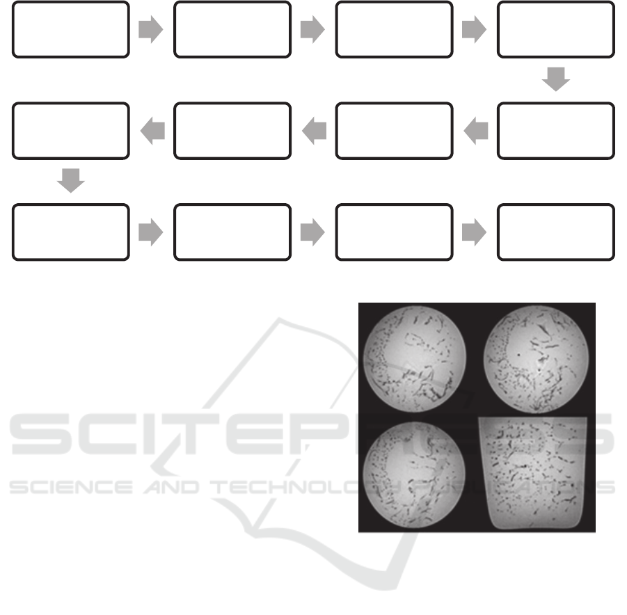
Figure 1: Process diagram for splint modeling.
the scan that produces a resonator requires a contrast
volume so that it can detect the sample since it would
not register something if it was introduced as is.
2.2 Magnetic Resonance Imaging
For this project a 7T MRI was used; each sample
scanned with a standard sequence weighted in T1
type Gradient-Echo in three-dimensional acquisition
(GE3D), with the parameters: TR 4 ms, TE 2.3 ms,
FA 20
o
, FOV 60x60 mm, MAT 256x256, slice 0.39
mm and Avg. 10 (Fig. 2). The studies acquired were
exported in DICOM format for post-processing.
2.3 The Software and 3D Printing
The MRI software consists basically of the OSIRIX
®
and AMIRA
®
programs, used in order to post-process
MRI (semi-automatic segmentation), generating the
Luffa model by polygonal approaches and exported to
STL format. The STL were processes with
Rhinoceros
®
CAD / CAM, its Grasshopper
™
plug-in,
Netfabb
®
by Autodesk and Stratasys Dimension
Elite
™
, thread fusion printer. The printing material
was thermoplastic ABS.
3 RESULTS AND DISCUSSION
An image obtained by a scanner such as the MRI is
made up of a point-cloud. The point-cloud is a set of
data collected by the scanner using the means in
three-dimensional structures. These points constitute
vertices in a three-dimensional coordinate field, so
they can be interpreted in a graphic format such as the
Figure 2: GE3D acquired in the 7T MRI scanner, without
post-processing.
STL the digital model is made up of multiple
polygons, which makes easy manipulate information
–as if it will be a physical object- in some program
such as Rhinoceros
®
(Fig. 3).
One of the images in STL was processed. First it
was increased by 1200% its size and debugging in the
Netfabb
®.
Next was printed in 3D (Fig. 4). As 3
rd
step,
verify its materialization and check its structure’s
behavior. The experiments worked quite well, so the
project continued, dissecting the virtual sample to
create a cubic module of approximately 15.6 cm
3
(Fig. 5), with which the splint structure could be
formed.
1)SAMPLING
• ObtainingsamplesoftheLuffa
specimen
•SamplepreparationforMRI
2)7TMRI
• Scanningofeachsample
• Postprocessingoftheimages
obtained
3)SELECTION
• Selectionofthebestimageby
structuralconformation
4)CONVERSION
•Conversionoftheselectedimage
toSTLformat
5)1st.PREPARATION
• Debuggingandcorrectingthe
imageinSTLwithNetfabb,for
printingtest
6)1st.PRINT
•STLmodelprintingtestinABS
plastic,tocheckprintingfeasibility
7)DISSECTION
• Digitaldissectionofapartofthe
imageinSTL,usingtheRhinoceros
8)CUBICMODULE
•Creationofthecubicmoduleusing
theRhinoceros
9)GEOMETRIZATION
• Geometryofthecubicmodule
usingtheGrasshopper
10)TESSELLATION
• Tessellationofthegeometrized
cubicmodule,onthesurfaceofa
humanfootmodeltocreatethe
splint,occupyingtheGrasshopper
11)2nd.PREPARATION
• Debuggingandcorrectingthesplint
model,withtheNetfabb program,
for3Dprinting
12)2nd.PRINT
•3DplasticprintingonABSscale
modelofthesplint
BIOIMAGING 2020 - 7th International Conference on Bioimaging
152
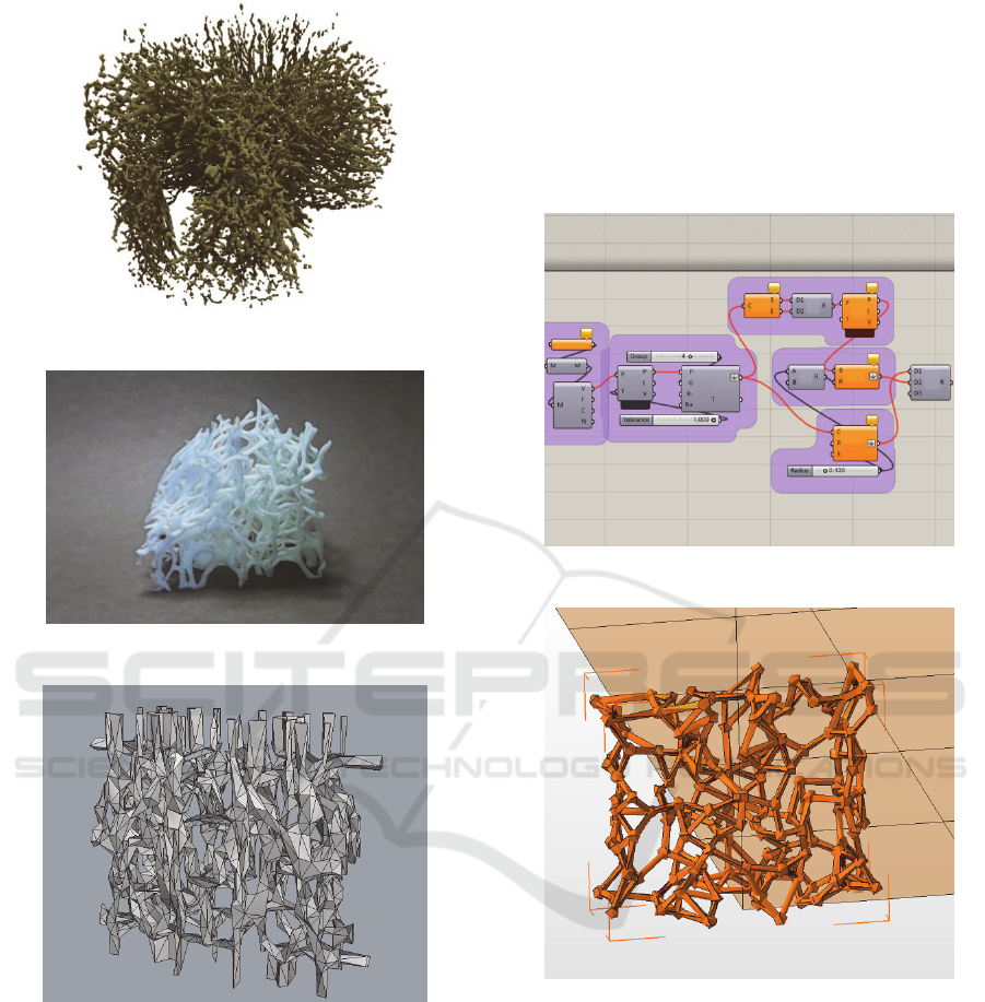
Figure 3: STL digital model of the Luffa sample.
Figure 4: 3D printing test on ABS plastic.
Figure 5: Dissected cubic module of the selected sample.
The dissected cubic module was made using
symmetry in the 3D planes, a section of the virtual
sample. In the splint modeling, was used the
Grasshopper
™
visual programming language that is
part of the Rhinoceros
®
program; this plug-in works
based on graphic algorithms called ‘variables’ with
which they can model and create shapes, through a set
of instructions called "definition".
To be able to work the cubic module, it was
necessary to convert it to geometric figures through
the Grasshopper
™
program, since images made based
on geometric figures are easier to handle due to their
low "weight", than those made of polygons. So a
“definition” was created for this purpose (Fig. 6) that
linked the "points" through cylinders coupled with
spheres, the result of which is seen in Figure 7. A
second “definition” (Fig. 8) was created to tessellate
the geometrized cubic module on the surface of a
virtual model of average human foot to generate the
splint.
Figure 6: “Definition” to geometrize the cubic module.
Figure 7: Geometrical cubic module.
“A tessellation is a special pattern, as it is formed
by different modules that splice together, according
to different symmetry principles, without overlapping
or leaving empty spaces” (Roncoroni Osio, 2017, p.
229).
Several dissected cubic modules accommodated
to determine which of them best covered the surface
(the distribution surface was the form of the human
foot), the optimum being 30 modules on the x-axis by
15 on the y-axis (fig. 9). It was observed that two
factors influence the formation of the splint a) the
dissected cubic module size and b) the size of the
distribution surface. For smaller modules, the mesh
3D Printed Human Foot Splint, Designed from MRI of the Luffa Cylindrica Dried Fruit
153
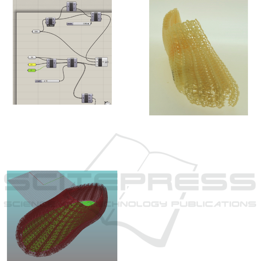
Figure 8: Second “definition” to tessellate the cubic
module.
becomes more closed and compact; on the contrary if
it is enlarged it becomes more open. The same goes
for the surface; more modules create a larger size of
the model and less for smaller. It is important to
mention that the surface where the module is
tessellated must be “closed”.
Figure 9: Result of the tessellation of the cubic module on
the foot model.
Next, it was exported to the Netfabb
®
program to
correct the file and prepare the model for ABS
printing, using the FDM process. Since the printer
used has a print volume of 8 in
3
, it was necessary to
reduce the dimensions of the splint by proportional
scaling so that the formal properties of the cubic
module was not lost so that it entered the printing tray,
in addition divided in half to appreciate the thickness
and arrangement it would acquire (fig. 10). The
printing process took 72 h, plus the removal of
soluble support material.
Figure 10: The First model of the foot splint, printed in ABS
plastic.
The results of this first phase were successful,
since the task of replicating the structure of the
organism as well as its application in the design of the
splint was achieved.
However, the work does not end here; it will
continue to improve in one case and resolve in
another, certain aspects such as the system of
attachment and coupling to the member, the final
material, the copy of the user's anthropometry and the
processing time. It is very important to compare the
three-dimensional model against the model printed by
FDM, since the first one exhibits a different behavior
from the second, especially due to the printing
trajectories of the filament, so that stress analysis and
resistance tests will also be done.
4 CONCLUSIONS
Bio-design, in conjunction with the NT –particularly
the MRI and 3D printing - is a good alternative in
product development, as demonstrated in this project.
Relying on principles backed by millions of years of
testing, in the great laboratory that is Nature, becomes
a great advantage when it comes to solving design
problems. Although the use of MRI is associated with
the medical area, it can be used equally for other
research; in this case it was used to obtain an exact
reproduction of the structure of a biological organism,
which otherwise would have been complicated to
achieve: the dried fruit of the Luffa. Its peculiar
structure gives it interesting mechanical properties
that make it resistant to deformation, which is why it
BIOIMAGING 2020 - 7th International Conference on Bioimaging
154

was thought that this advantage could be used to
design a splint for the human foot, which improves on
the traditional ones made of coated bandages with
plaster This research can serve as a basis for creating
diverse and varied bio-designed products, not only
from the medical area, but also from other fields and
specialties of human activity.
ACKNOWLEDGMENTS
We thank the National Center for Research in
Medical Imaging and Instrumentation (CI
3
M) of the
Universidad Autónoma Metropolitana, Iztapalapa
Unit, for the loan of the 7T MRI scanner and the use
of its facilities, as well as the Materialization and 3D
Modeling Laboratory of the Universidad Autónoma
Metropolitana Azcapotzalco Unit for the realization
of this project. Thanks to the Librarian César
Saavedra-Alamillas, for his help in the bibliographic
review.
REFERENCES
Al Jabbari, O., Abu Saleh, W. K., Patel, A. P., Igo, S. R., &
Reardon, M. J. (2016). Use of three-dimensional
models to assist in the resection of malignant cardiac
tumors. Journal of Car-diac Surgery, 31(9), 581–583.
https://doi.org/10.1111/jocs.12812
Backstrom, K., Nazari, M., Gu, I. Y.-H., & Jakola, A. S.
(2018). An efficient 3D deep convolutional network for
Alzheimer’s disease diagnosis using MR images. 2018
IEEE 15th International Symposium on Biomedical
Imaging (ISBI 2018), 149–153. https://doi.org/
10.1109/ISBI.2018.8363543
Bezinque, A., Moriarity, A., Farrell, C., Peabody, H.,
Noyes, S. L., & Lane, B. R. (2018). Determination of
Prostate Volume. Academic Radiology, 25(12), 1582–
1587. https://doi.org/10.1016/j.acra.2018.03.014
Bouchet, A., Pastore, J. I., Brun, M., & Ballarin, V. (2015).
Segmentation of Lateral Ventricles in Magnetic
Resonance Images. En A. Braidot & A. Hadad (Eds.),
VI Latin American Congress on Biomedical
Engineering CLAIB 2014, Paraná, Argentina 29, 30 &
31 October 2014 (Vol. 49, pp. 457–460).
https://doi.org/10.1007/978-3-319-13117-7_117
Chen, Q., Shi, Q., Gorb, S. N., & Li, Z. (2014). A multiscale
study on the structural and mechanical properties of the
luffa sponge from Luffa cylindrica plant. Journal of
Biomechanics, 47(6), 1332–1339. https://doi.org/
10.1016/j.jbiomech.2014.02.010
Cole-Hamilton, D. J., Kaye, B., Chudek, J. A., & Hunter,
G. (1995). Nuclear Magnetic Resonance Imaging of
Waterlogged Wood. Studies in Conservation, 40(1), 41.
https://doi.org/10.2307/1506610
Ehricke, H.-H., Hauser, T.-K., Nägele, T., Schult, T., &
Klose, U. (2018). Magnetic resonance angiography
contrast enhancement and combined 3D visualization
of cerebral vasculature and white matter pathways.
Computerized Medical Imaging and Graphics, 70, 29–
42. https://doi.org/10.1016/j.compmedimag.
2018.09.005
Ghisalberti, E. L., & Godfrey, I. M. (1998). Application of
nuclear magnetic resonance spectroscopy to the
analysis of organic archaeological materials. Studies in
Conservation, 43(4), 215–230. https://doi.org/10.1179/
sic.1998.43.4.215
Kim, H., & Jeong, S. (2015). Case study: Hybrid model for
the customized wrist orthosis using 3D printing.
Journal of Mechanical Science and Technology,
29(12), 5151–5156. https://doi.org/10.1007/s12206-
015-1115-9
Pal, A. (2016, julio 25). Orthopedic Splints—Principles,
Types and Use. Retrieved on September 19, 2019, from
Bone and Spine website: https://boneandspine.com/
orthopedic-splints/
Pawlyn, M. (2011). Biomimicry in architecture (1th
edition). London: Riba Publishing.
PROTA. (2018). Luffa cylindrica. Retrieved on March 19,
2019, from https://www.prota4u.org/database/protav8
.asp?h=M4&t=Luffa,cylindrica&p=Luffa+cylindrica#
Synonyms
Roncoroni Osio, U. (2017). Manual de diseño generativo.
Retrieved from https://ra.ocls.ca/ra/algologin.aspx?inst
=algonquin&url=http://search.ebscohost.com/login.asp
x?direct=true&scope=site&db=nlebk&AN=1601346
Shen, J., Min Xie, Y., Huang, X., Zhou, S., & Ruan, D.
(2012). Mechanical properties of luffa sponge. Journal
of the Mechanical Behavior of Biomedical Materials,
15, 141–152. https://doi.org/10.1016/j.jmbbm.
2012.07.004.
3D Printed Human Foot Splint, Designed from MRI of the Luffa Cylindrica Dried Fruit
155
