Femur Fracture Detection Based on Deep Learning Model YOLOv8
Dongru Xie, Hongjian Yu
*
, Zhijiang Du, Hao Wang, Xiangyu Shen and Zhenyi Wang
State Key Laboratory of Robotics and System, Harbin Institute of Technology, Harbin, China
Keywords: Femur Fracture, X-Ray Images, Object Detection, Image Pre-Process, Deep Learning.
Abstract: Femur fracture occurs in various circumstances like car accidents, high-altitude fall incidents, tumour illness,
and elderly falls. For better recognition and treatment, physicians need to search the X-ray images for
fracture detail. However, some X-ray images were unclear to diagnose, and some were taken from the side
position, which is difficult to detect the fracture. This study uses the YOLOv8 model to help physicians with
femur fracture detection by utilizing deep-learning models. The performance of YOLOv8 is 42.35% in
AP50:95, 84.24% in mAP50, and 25.45% in mAP75 on the private dataset is from Shenzhen University
General Hospital. The result shows that the YOLOv8 detection model is competitive and faster on the
personal femur fracture dataset than YOLOv3 and YOLOv5 models.
1
INTRODUCTION
Bone fractures are regular in hospitals due to car
accidents, high-altitude fall incidents, tumor illness,
and elderly falls. Physicians use medical images, for
example, X-ray images, to search for the detail of
the fracture. However, due to the angle of images
taken can vastly alter the fracture info, the deep
learning model YOLOv8 is used to detect femur
fracture.
YOLOv8 is a multi-scale detection model. It uses
three detection heads to classify. According to the
basic of the deep learning model, YOLOv8 can learn
simple features like straight lines and oblique lines
at a low level. At a high level, it can learn more
complex features like the femur, femur shaft, and
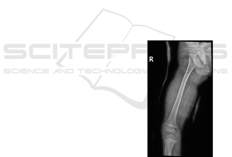
femoral head. Figure 1 shows the anatomy of the
femur. The fracture can occur at the femoral shaft,
femoral distal, and femoral proximal.
Femoral proximal mainly include the femoral
neck, intertrochanteric, and subtrochanteric femoral
fractures. Femoral neck fractures occur
predominantly in the elderly, typically resulting
from low-energy falls, and may be associated with
osteoporosis.
Femoral shaft fractures are among the most
common fractures seen in orthopedic practice. The
femur is the most prominent bone in the body and
one of the primary weight-bearing bones of the
lower limbs, and unless treated appropriately,
fractures can lead to long-term morbidity and
disability.
Distal Femur often is unstable and comminuted
and tends to have a bimodal distribution, occurring
in elderly or younger multiple-injured patients.
Figure 1: The anatomy of the femur.
This research used the YOLOv8 model to detect
the fracture for better detection. This deep learning
model has three detection heads, which are 8x, 16x,
and 32x. Different scales can detect different scales
of objects. For instance, if 8x can detect the femur,
then 16x can detect the approximate location where
190
Xie, D., Yu, H., Du, Z., Wang, H., Shen, X. and Wang, Z.
Femur Fracture Detection Based on Deep Learning Model YOLOv8.
DOI: 10.5220/0012277300003807
Paper published under CC license (CC BY-NC-ND 4.0)
In Proceedings of the 2nd International Seminar on Artificial Intelligence, Networking and Information Technology (ANIT 2023), pages 190-193
ISBN: 978-989-758-677-4
Proceedings Copyright © 2024 by SCITEPRESS – Science and Technology Publications, Lda.

the femur is, and 32x can use the bounding box to
locate the fracture.
The accuracy score obtained from the fracture
detection performed by Rashid et al., using a 28-
layer dilated CNN and long short-term memory
(DCNN-LSTM) on 965 wrist X-ray images, is
88.24% (Rashid, 2023). The result of fracture
detection performed by Jia et al. on 1227 sternum
fracture X-ray images from the collection of sternal
radiographs and hospital diagnostic reports, 0.71
mAP, was obtained using the cascade R- CNN
method (Jia, 2022). The AP score of Guan et al. was
62.04% with a two-stage R-CNN method developed
for fracture detection based on nearly 4000 arm
fracture X-ray images using Resnet backbone.[Guan
B, 2020] Wang et al. carried out fracture detection
procedures(WrisNet), achieving a 56.6% score of
AP, using the model inspired by Faster-RCNN,
mainly composed of ResNeXt-TA and FPN for a
total of 4346 hairline fractures in hand X-rays
images.(WANG W.) Lu et al. developed automated
universal fractures detection in X-ray images using a
modified Ada-ResNeSt backbone network and the
AC-BiFPN detection method based on the part of
the MURA dataset. They achieved an AP score of
68.4% on 30000 X-ray images. (LU S, 2022) Guan
et al. achieved an AP score of 88.9% using a
balanced FPN-ResNeXt model developed for
fracture detection in a 3842 thighbone X-ray
radiographs dataset. (Guan B, 2022) Yadav et al.
used a deep learning model to detect and classify X-
ray images of human fracture bone and healthy bone.
5-fold cross-validation was implemented on 4000
augmented datasets and got 92.44 % accuracy for
the healthy and the fractured bone. (Xue L, 2021)
ParallelNet is proposed by Wang et al. for detection
tasks on thigh bone fracture based on multiple
backbone networks. The dataset contains 3842 X-
way radiographs; the result is 87.8% AP50 and
49.3% AP75. (WANG M, 2021) Chin et al.
proposed an Auxiliary Classifier Generative
Adversarial Network (AC-GAN) model to label the
position of the fracture. The result shows an
accuracy of 91.2% (Chiun-Li Chin, 2019).
2
ANOTHER SECTION OF YOUR
PAPER
YOLOv8 is the latest deep-learning model in the
YOLO series. The structure of this model is shown
in Figure 2. The detail of the model will be
explained in this section.
Downsample 4X
Downsample 16X
Downsample 8X
Downsample 32X
Downsample 16X
Down sample 8X
Downsample 16X
Downsample 32X
C2f & ConvModule
C2f & ConvModule
C2f & SPPF
Upsample & Concat
C2f & Upsample & Concat
C2f
2x ConvModule
Femur
Frac ture
C2f & ConvModule
Head1
ConvModule
Head2
Head 3
BACKBO NE BODY
Figure 2: The structure of YOLOv8 model.
2.1 Yolov8 Model
Like YOLOv5, YOLOv8 provides different size of
models based on the scaling factor to meet the needs
of different scenarios. The Backbone and Neck part
of the model refers to the YOLOv7 ELAN design
idea. The C3 structure from YOLOv5 has been
replaced with a richer C2f structure of gradient flow,
and the number of channels has been adjusted for
models of different scales. It is a fine-tuning of the
model structure. It is no longer a brainless set of
parameters to apply to all models, dramatically
improving the model's performance. Compared with
YOLOv5, the Head part has changed a lot. It has
been replaced with the current mainstream
decoupling head structure, which separates the
classification and detection heads. The
TaskAlignedAssigner positive sample allocation
strategy is adopted in the calculation, and the data
enhancement part of the Distribution Focal Loss
training is introduced. The Mosiac enhanced
operation can effectively improve the accuracy
introduced from YOLOX.
2.2 Study Dataset
The X-ray images of femur fracture include 312
fracture images collected from Shenzhen University
General Hospital between 2019 to 2023, ranging
from femoral neck fracture to distal femur fracture.
The physicians from Shenzhen University General
Hospital checked the data labeling of femur X-ray
images.
3
EXPERIMENTS
This study used local PC to train the deep-learning
model for femur fracture detection. The graphics
card of the local PC is 12 GB Nvidia GTX3060.
The following configuration is used in all
machine learning models for femur fracture
detection: the epoch of data training is 300 times, the
Femur Fracture Detection Based on Deep Learning Model YOLOv8
191
initial learning rate is set as 0.01, and the final
learning rate is 0.01 of the initial learning rate.
Warmup epochs are 3. The parameters used to
optimizer weight decay are 5e-4, initial warmup
momentum is 0.8.
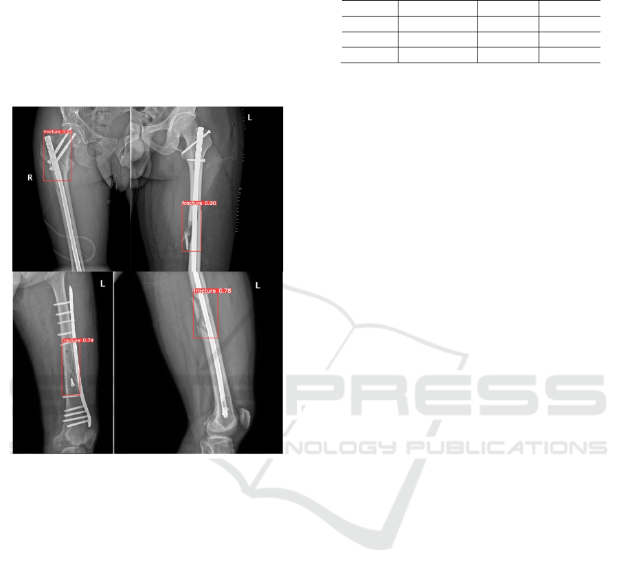
The bounding box outputs performed with the
YOLOv8 model in femur fracture X-ray images are
provided as a dataset sample in Figure 3 below.
Figure 3: Sample of Detection result of YOLOv8 model.
In order to explain the detection result of
YOLOv8, this research adds other deep learning
models to compare the result of YOLOv8.
YOLOv3 uses more profound and more accurate
Darknet-53 as the backbone and shifts from multi-
category to multi-label classification, removing
softmax and using binary cross entropy instead.
The network architecture of YOLOv5 consists of
three parts: CSPDarknet as the backbone, PANet as
the neck, and Yolo Layer Head. The data is first
input to CSPDarknet for feature extraction and then
input to PANet for feature fusion. Finally, the Yolo
layer outputs the detection results.
Based on the same parameters, the detection result
of three deep learning models is shown in the table
below.
Table 1: Detection result of YOLOv8, YOLOv5, and
YOLOv3 models.
Model mAP50-95 mAP50 mAP75
YOLOv8 0.4235 0.8424 0.2545
YOLOv5 0.3627 0.7578 0.3146
YOLOv3 0.4079 0.8116 0.3666
Table 1 shows that YOLOv8 has better overall
detection results than YOLOv5 and YOLOv3
Models on fracture detection. When the results
mentioned above are examined, it depicts that
YOLOv8 has an improvement in mAP50-95 and
mAP50 values. YOLOv8 can be used in femur
fracture detection in the private dataset.
4
CONCLUSION
This study aims to support physicians and intern
doctors in medical image detection and solve the
problem when X-ray images are deficient in clinical
needs and require physicians to retake X-rays of
femur fractures. In addition, the result shows that the
YOLOv8 model can detect femur fracture better
than other deep-learning models.
ACKNOWLEDGMENTS
This work was financially supported by Key-Area
Research and Development Program of Guangdong
Province (No.2020B0909020002) and Self-Planned
Task (No.SKLRS202211B) of State Key Laboratory
of Robotics and System (HIT).
REFERENCES
Rashid T, Zia M S, Najam UR R, et al. A Minority Class
Balanced Approach Using the DCNN-LSTM Method
to Detect Human Wrist Fracture [J]. Life-Basel, 2023,
13(1). https://doi.org/10.3390/life130 10133
Jia Y, Wang H, Chen W, et al. An attention-based cascade
R-CNN model for sternum fracture detection in X-ray
images [J]. CAAI Transactions on Intelligence
Technology, 2022, 7(4): 658-70. https://doi.org/
10.1049/cit2.12072
Guan B, Zhang G, Yao J, et al. Arm fracture detection in
X-rays based on improved deep convolutional neural
network [J]. Computers & Electrical Engineering,
2020, 81. https://doi.org/ 10.1016/j.compeleceng.2019
.106530
Wang W, Huang W, Lu Q, et al. Attention mechanism-
based deep learning method for hairline fracture
detection in hand X-rays [J]. Neural Computing &
ANIT 2023 - The International Seminar on Artificial Intelligence, Networking and Information Technology
192

Applications, 2022, 34(21): 18773-85. https://
doi.org/10.1007/s00521-022-07412-0
Lu S, Wang S, Wang G. Automated universal fractures
detection in X-ray images based on deep learning
approach [J]. Multimedia Tools and Applications,
2022, 81(30): 44487-503. https://doi.org/10.1007/
s11042-022-13287-z
Guan B, Yao J, Wang S, et al. Automatic detection and
localization of thighbone fractures in X-ray based on
improved deep learning method [J]. Computer Vision
and Image Understanding, 2022, 216.
https://doi.org/10.1016/j.cviu.2021.103345
Xue L, Yan W, Luo P, et al. Detection and localization of
hand fractures based on GA_Faster R-CNN [J].
Alexandria Engineering Journal, 2021, 60(5): 4555-
62. https://doi.org/10.1016/j.aej.2021.03.005
Wang M, Yao J, Zhang G, et al. ParallelNet: multiple
backbone network for detection tasks on thigh bone
fracture [J]. Multimedia Systems, 2021, 27(6): 1091-
100. https://doi.org/10.1007/s00530-021-00783-9
Chiun-Li Chin, Yong-Long Lin, Yu-Chieh Liu. Various
Types Fracture Labeling In Bone Radiographs Using
Modified AC-GAN[C], Proceedings of the
International Conference on Technologies and
Applications of Artficial Intelligence (TAAI),
Kaohsiung, TAIWAN, 2019 Nov. 21-23.
https://doi.org/10.1109/TAAI48200.2019.8959863
Femur Fracture Detection Based on Deep Learning Model YOLOv8
193
