A Comprehensive Analysis of Medical Image Fusion Techniques:
A Detailed Review
T. M. Hayat
1,*
and Sai Madhavi D.
2,†
1
Ballari Institute of Technology and Management, Ballari, Visvesvaraya Technological University, Belagavi-590018, India
2
RaoBahadur Y Mahabaleshwarappa Engineering College, Ballari, Visvesvaraya Technological University,
Belagavi-590018, India
Keywords: CT, Image Fusion, Image Processing, MRI.
Abstract: Image fusion involves merging a collection of images of the same scene to create a single composite image.
Its purpose is to generate a more visually appealing image or to extract additional valuable information from
it. The main aim of image fusion is to produce a new image that contains high-quality data, which cannot be
obtained through other means. This process combines multisensor, multiview, and multitemporal data to
create a single, comprehensive image. Image fusion techniques have been applied in various fields, including
remote sensing, astronomy, and medical imaging. In medical imaging, image fusion has been particularly
useful for simultaneously evaluating CT, MRI, and PET images to find what type of disease or its effect. In
this paper, we present a novel literature review on image fusion techniques applied to medical images. Our
findings suggest that image fusion can greatly improve the clinical reliability of disease diagnosis and
analysis, and we anticipate strong growth in this field in the near future.
1 INTRODUCTION
Image fusion is a progression method of merging a set
of images of the same scene into one composite
image. Fusioning is done in order to get an enhanced
image or to enhance some useful information from it.
Image fusion in medical field has seen significant
growth several years and it incorporates a broad range
of techniques in the field of image fusioning.
The fusion process aims to address medical
conditions or diseases by analyzing images of the
human body, organs, and cells. With advancements in
computer-aided imaging techniques, this process
helps medical experts make more informed decisions
in a shorter amount of time. By using fusion methods
on multi-sensor and multi-source images, a wider
range of features can be used for medical analysis
which is leading to more precise information
processing and the ability to uncover details that may
be invisible to the human eye. Additional Information
is obtained from fused images, which lead to locate
abnormalities, more accurately. Image filters are
indeed a fundamental concept in image processing,
*
Research Scholar
†
HOD and Prof
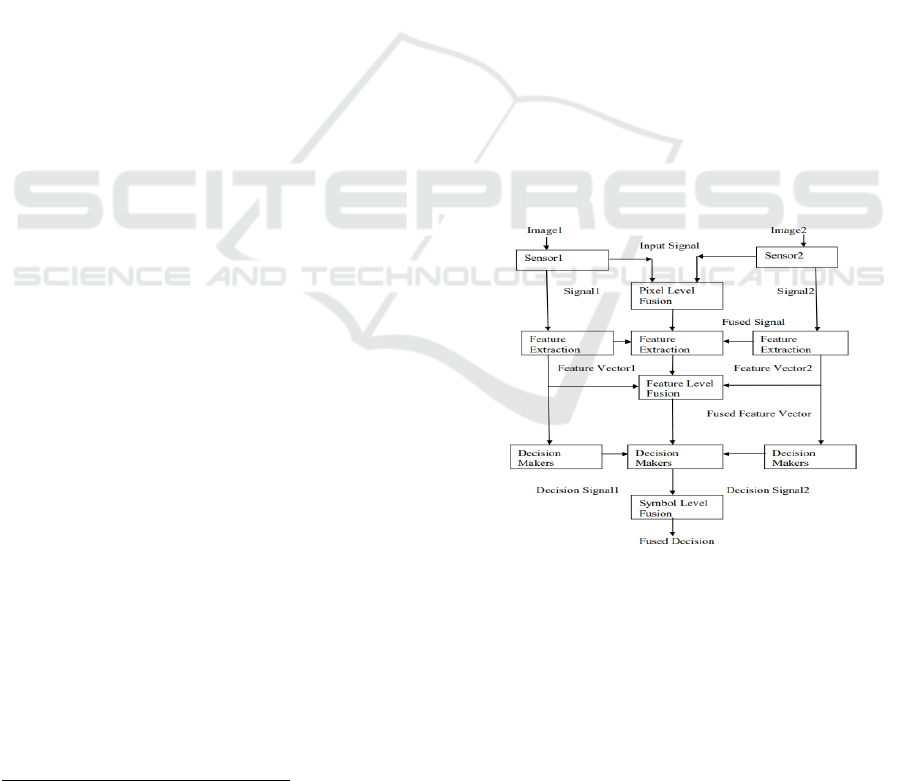
Figure 1: Information fusion system at all three levels of
processing.
and they are used to enhance, manipulate, or extract
information from images. There are various types of
image filters, such as spatial filters, frequency filters,
and edge detection filters, each serving a specific
purpose in image processing. In spatial domain
filtering, a filter is applied directly to the pixels of the
Hayat, T. and Madhavi D., S.
A Comprehensive Analysis of Medical Image Fusion Techniques: A Detailed Review.
DOI: 10.5220/0012603200003739
Paper published under CC license (CC BY-NC-ND 4.0)
In Proceedings of the 1st International Conference on Artificial Intelligence for Internet of Things: Accelerating Innovation in Industry and Consumer Electronics (AI4IoT 2023), pages 147-152
ISBN: 978-989-758-661-3
Proceedings Copyright © 2024 by SCITEPRESS – Science and Technology Publications, Lda.
147
image, and it can be utilized for smooth or sharpen
images, remove noise or enhance specific features.
Spatial filters are often used for real-time image
processing applications, such as video or camera
feeds, as they are faster than frequency domain filters.
Image processing has various applications, as you
have mentioned. It is used in computer vision tasks,
such as object recognition, face detection, and motion
tracking. In security, image processing can be used
for surveillance systems and access control. It is also
used in entertainment and gaming industries, for
creating special effects or developing interactive
games. One of the most important uses of image
processing is in medical imaging. Medical images,
such as X-rays, CT scans, or MRI, are used to
diagnose and treat various diseases and conditions.
Image processing can help improve the accuracy of
medical image analysis, by enhancing the contrast
between different tissues, segmenting specific
regions of interest, or detecting abnormalities.
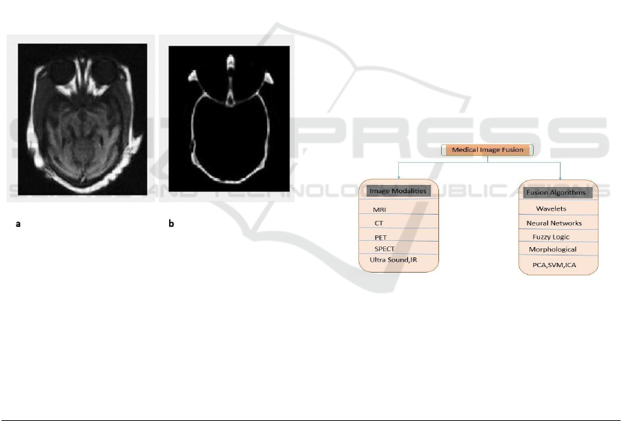
Figure 2: (a) MRI Image used as source image (b) CT
Image.
Deep learning models can also be trained on huge set
of database of medical images, to automatically
detect and diagnose certain conditions, such as cancer
or fractures. This can greatly improve the speed and
accuracy of medical diagnosis, and help doctors make
more informed decisions about patient care.
Medical Image Fusion.
Medical image fusion comprises of processing and
grouping of multiple images acquired from single or
multiple imaging modalities. The key aim of this
process are to increase the quality of medical images,
decrease randomness and redundancy, and increase
their clinical applicability for diagnosis and
evaluation of medical problems. Several multimodal
medical image fusion algorithms and devices have
shown great progress in improving the accuracy of
clinical decisions based on medical images. This
overview classifies the process of fusioning of
medical image research based on his three factors:
(a) commonly used image fusion methods,
(b) relevant imaging modalities;
(c) Organ examined.
Despite numerous open technical and scientific
challenges, medical image fusion process has shown
encouraging results in augmenting the clinical
dependability of medical images for diagnostics as
well as analysis. It is a rapidly growing scientific area
with the potential for significant advancements in the
future years The fusion of medical images controls
the noteworthy and complementary information of
various images those are retrieve from the different
sources that used for identify the diseases and better
treatment.
Figure 3: Modalities and Algorithms of image fusion
studies.
The field of medical image analysis is distributed
into six different categories as depicted below:
Table 1: Categories of Medical Image Analysis.
Categories
Description
Post-acquisition
Prior to diagnosis, images are often subjected to preprocessing techniques such as denoising and
renovation to improve their quality and make them usable.
Segmentation
The accurate diagnosis of medical images such as CT scans of abdomen or MRI scans of brain
which requires the identification and definition of important features, such as organs, within
the image. This process, known as delineation, is crucial for effective analysis and interpretation.
Registration
The process of registering or aligning captured images with a model or previous image is a
crucial requirement in computer-assisted surgery.
AI4IoT 2023 - First International Conference on Artificial Intelligence for Internet of things (AI4IOT): Accelerating Innovation in Industry
and Consumer Electronics
148

Computation
In various computer-assisted therapies, there is also a need for the calculation of physical
quantities and the execution of additional computational tasks such as fusion and compression.
Visualization
It is crucial for medical images to be displayed, so that medical professionals can accurately
diagnose diseases.
Security
Personal medical health information is highly sensitive and must be properly secured through
methods such as watermarking. This ensures that only legal users have access to the information
and that it is accurately linked to the correct medical record for the appropriate patient.
Medical image fusion can be categorized as shown
below: -
1.) Multi View Image Fusion: In this fusion
images are taken from different viewpoints but have
same modality and at the same time.
2.) Multi Modal Image Fusion: In this fusion
different sensors like CT, MRI and PET etc. are used
to collect the images (IJARIIE, n.d.). Clinical
precision are improve by using the Multimodal
medical image fusion algorithms and devices.
3.) Multi Temporal Image Fusion: In this type of
fusion, images are extracting at different times for
finding the changes between images.
4.) Multi Focus Image Fusion: In this type of
fusion images are taken from a 3D scene continually
with various focal lengths (Mishra and Bhatnagar,
2014 ).
2 LITERATURE REVIEW ON
MEDICAL IMAGES
P. James and B.V. Dasarathy (James and Dasarathy,
2014) stated that multimodal fusion of medical
images has shown significant improvement in clinical
diagnosis of disease. Fusion using Multimodel of
medical images has shown great improvement in
clinical diagnosis of disease. This review article
provides practical techniques and summarizes the
challenges in medical image fusion. In this white
paper, while there are numerous scientific challenges
and open technologies available, medical image
fusion is a highly useful technology for improving
clinical consistency, identification, and analysis.
Daniel Ruijters (Ambrosini et al., 2017) proposed
that medical scanning technology gives a wide
spectrum of valuable and harmonizing information
about a patient's physiology, anatomy and pathology,
but the optimal exploitation of this wealth of
information is a tough job.
Deron Rodrigues et al., (Rodrigues et al., 2014)
have explained that image fusion has become
important part in medical field for diagnosis or
analysis of disease. The paper describes the
introduction of image fusion methods by using
wavelet transform and the comparison between the
performance of the various types of wavelet basis
families used.
Hari Om Shanker Mishra and Smriti Bhatnagar
(Mishra and Bhatnagar, 2014) have explained that
fusing techniques are used for image enhancement in
several imaging techniques like Computed
Tomography (CT) and Magnetic Resonance Imaging
(MRI).
Hiral Rameshbhai Patel and Raviraj Chauhan
(IJARIIE, n.d.) proposed a decomposition and
reconstruction method to improve image quality. The
Discrete Ripplet Transform is an advanced
directionality and localization transform for such
edges, and the combination of DWT and DRT yields
better images than DWT.
Jan Flusser et al. (Zitova and Flusser, 2003),
stated as fusion process of various images is used in
numerous applications, such as astronomy, multi-
sensor fusion, medical imaging, military, remote
sensing, security and surveillance fields. used in these
applications. Image fusion is used in many
applications such as remote sensing and medical
fields, and this pattern is primarily used in CT and
MRI images where the more accurate the image, the
more useful the information. Many approaches have
been developed for medical image fusion.
M.D. Nandeesh and Dr. M. Meenakshi (Casey
and Damper, 2010) studied about image fusion
techniques with their performance evaluation
analysis. They used Discrete Wavelet Transform,
Curvelet Transform, Principle Component Analysis,
Stationary Wavelet Transform techniques etc.
Madhusmita Sahoo (Ambrosini et al., 2017)
explains a modern fusion method to enhance the
information content of the fused image. The
technique uses wavelet transform, maximum
selection rule, windowing technique and GLCM
based segmentation.
Mc Cassey et, al. (Casey and Damper, 2010) uses
image fusion algorithm to acquire the sincere feasible
depth-of-field in macro-photography by using typical
digital camera images. Macro photography has some
primary problems, one of the most critical is the
difficulty of insufficient lighting. Mayank Agrawal et
al. (Agrawal et al., 2010) proposed a fusion algorithm
A Comprehensive Analysis of Medical Image Fusion Techniques: A Detailed Review
149
for multispectral magnetic resonance imaging that
preserves both component and edge information and
provides better performance associated to existing
fusion algorithms.
Medha Balachandra Mule and Padmavathi N.B
(Kotian et al., n.d.) have done analysis of different
medical imaging modalities used in fusion. They have
explained and compared different image fusion
techniques using the quality metrics Peak Signal to
Noise Ratio (PSNR) and Root Mean Square Error
(RMSE).
Nayera Nahvi and Deep Mittal (Niranjan and
Patel, n.d.) have explained a new algorithm for
multimodal medical image fusion based on DWT
technique. The algorithm escalates the quality of
multimodality medical image fusion and the output
reveal the efficiency of fusioning process.
P.Ambika Priyadharsini and M.R. Mahalakshmi
(Priyadharsini et al., n.d. ) proposed that SVD is a
substitute image fusion method, which improves the
content of medical images by merging two or more
multimodal medical images.
Paul Hill et al. proposed DT-CWT (Hill et al.,
2005) techniques for image fusion in remote sensing,
robotics and medical applications. This method gives
better qualitative and quantitative output compared to
previous wavelet fusion techniques.
Periyavattam Shanmugam Gomathi and
Bhuvanesh Kalaavathi (Gomathi and Kalaavathi,
2016) states a comparative study of image fusion of
MRI and CT images based on various wavelets
transforms techniques is performed. The final fused
image is tested by using many performance metrics to
evaluate which wavelet gives the best output.A new
generation of high resolution satellite images with
less than 1 meter spatial resolution in panchromatic
mode is now available. This paper compares the
output of three different techniques to fuse the
multispectral information and panchromatic data of
Quick Bird satellite imagery.
Richa Singh et al. (Lawson et al., 2020) proposed
a fusion algorithm that uses Redundant Discrete
Wavelet Transforms to combine pairs of
multispectral magnetic resonance imaging such as
Proton Density, T2 and T1 brain images.
Helonde and Prof. M.R. Josh (Holende et al.,
2010) explained that image fusion plays an important
role in digital image reconstruction as re-processing
steps. Medical image fusion helps in easy diagnostics
and reduces the time gap between the diagnosis of the
disease and the treatment.
Dr. S. Manikanda Prabu et al. (Prabu and
Ayyasamy, 2014) used Lifting Wavelet Transform
(LWT) based three different medical image fusion
approaches and performed comparative analysis. The
IAV method was found to be more suitable for
medical image fusion than other approaches in
wavelet domain.
Walid Aribi et al. (Arabi et al., 2012), developed
new methods to evaluate the quality of medical
images based on the multi resolution fusion. These
methods are evaluated by objective technical quality.
Zhi-haiXu et al. (Jing, 2009) proposed a new
fusion algorithm based on wavelet transform by
analyzing the three fusion operators. The algorithm
was validated by CT/PET images.
Zhijun Wang et al. (Wang et al., 2005) presented
a complete outline of the General Image Fusion (GIF)
method is a useful framework for categorizing and
evaluating image fusion methods. One such method
is MRAIM, which stands for Multiresolution
Analysis-based Image Fusion using Morphological
Reconstruction and Iterative Method.
The field of Image-Guided Therapy (IGT) is
rapidly growing and has seen success with the
commercialization of advanced IGT systems by
several small companies. However, in meetings
between IGT investigators, it was determined that
there are several key areas that require collaborative
effort from the community to improve patient care.
Image fusion is an interesting field for the
researcher. Various techniques like wavelet transform
HIS and PCA based methods are proposed by many
author or researchers.
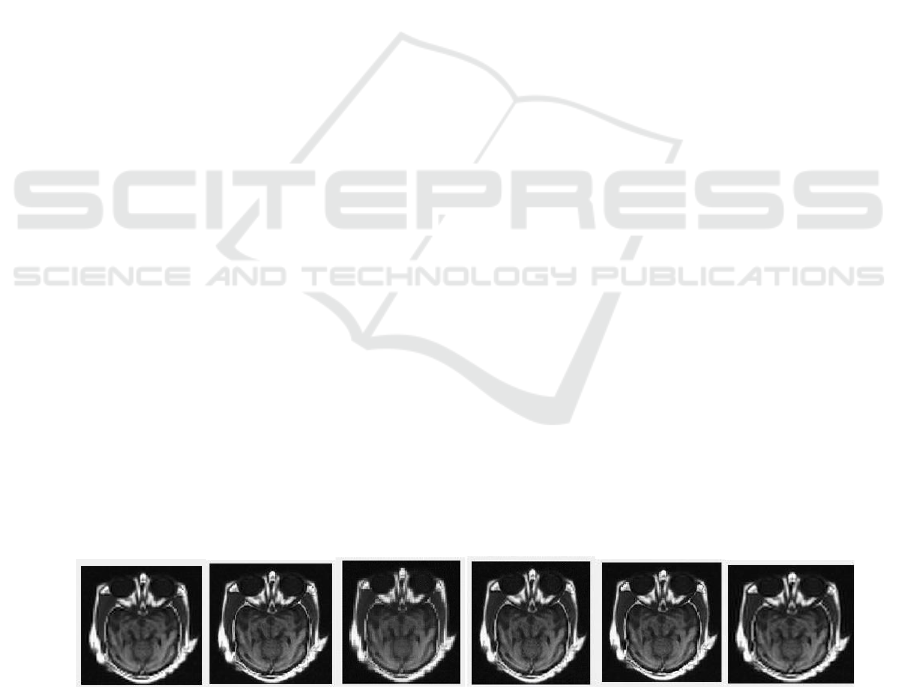
Figure 4: Fusion of MRI and CT Image a) Using Daubechies(db) b) Using Coiflets (coif)c) using Bi-orthogonal (bior) d) By
Symlets (sym) (e) By Reverse Bior (rbio) (f)By Discrete Meyer (dmey).
AI4IoT 2023 - First International Conference on Artificial Intelligence for Internet of things (AI4IOT): Accelerating Innovation in Industry
and Consumer Electronics
150

Table 2: Different Fusion Strategies.
METHOD
TYPE OF IMAGE
FUSION STRATEGIES
MORPHOL
OGY
KNOWLE
DGE
MRI, CT, ULTRA SOUND,
MAMMOGRAM, PET
MORPHOLOGY FILTERA,
LEARNING SYSTEMS,
EXPERT SYSTEMS
WAVELET
S
CT, PET, MRI, ULTRA
SOUND, SPECT
DISCRETE WAVELET TRANSFORMS,
STATIONARY WAVELET, MULTI-
WAVELET TRANSFORM
ANN
CT, PET, MRI, ULTRA
SOUND, MRA, SPECT
NEURAL NETWORKS, CLUSTERING
NEURAL NETWORKS
FUZZY
LOGIC
CT, PET, MRI, ULTRA
SOUND, MRA, SPECT
IMAGE FUZZIFICATION,
DEFUZZIFICATION, NEURO FUZZY
NETWORKS,
3 CONCLUSION
The field of medical diagnostics and monitoring is
rapidly advancing with the growth of latest
technologies and scientific advancements. However,
the use of medical images to aid in these processes is
not without challenges. These challenges can be
technological, scientific, and societal in nature.
One of the challenges is related to the quality of
imaging features. In order to achieve a
comprehensive understanding of a medical condition,
multiple imaging modalities are often used. However,
these modalities may produce images with different
qualities and characteristics. Image fusion techniques
can be used to improve the quality of imaging features
by integrating information from multiple modalities.
However, the key challenge in applying image
fusion algorithms to medical images is to confirm that
the medical relevance is maintained and that they aid
in achieving enhanced clinical outcomes. This
requires careful consideration of the specific medical
application, as well as the imaging techniques used.
Despite these challenges, image fusion techniques
hold great promise for improving the quality of
medical imaging and aiding in diagnostics and
monitoring of medical conditions. As such, ongoing
research in this area is critical for the advancement of
medical science and for the betterment of patient care.
REFERENCES
Wang, Z., Ziou, D., Armenakis, C., Li, D., & Li, Q. (2005).
A comparative analysis of image fusion methods. IEEE
transactions on geoscience and remote sensing, 43(6),
1391-1402.
Prabu, S. M., & Ayyasamy, S. (2014). An efficient
watermarking algorithm based on DWT and FFT
approach. International Journal on Computer Science
and Engineering, 6(6), 211.
Zitova, B., & Flusser, J. (2003). Image registration
methods: a survey. Image and vision computing,
21(11), 977-1000.
http://ijariie.com/AdminUploadPdf/Medical_Image_Fusio
n_Using_Combine_Approach_of_DWT_and_DRT_ija
riie2635.pdf
Holende et al., A special issue on biologically inspired
information fusion, Inform. Fus. 11 (1) (2010) 1.
M.C. Casey, R.I. Damper, Editorial: special issue on
biologically-inspired information fusion, Inform. Fus.
11 (1) (2010) 2–3.
Aribi, W., Khalfallah, A., Bouhlel, M. S., & Elkadri, N.
(2012, March). Evaluation of image fusion techniques
in nuclear medicine. In 2012 6th International
Conference on Sciences of Electronics, Technologies of
Information and Telecommunications (SETIT) (pp.
875-880). IEEE.
Jing, L. (2009). Scrambling analysis of ciliates (Doctoral
dissertation, Master’s thesis, University of
Saskatchewan, Saskatoon).
J. Greensmith, U. Aickelin, G. Tedesco, Information fusion
for anomaly detection with the dendritic cell algorithm,
Inform. Fus. 11 (1) (2010) 21–34.
Lawson, K. A., Sousa, C. M., Zhang, X., Kim, E., Akthar,
R., Caumanns, J. J., ... & Moffat, J. (2020). Functional
genomic landscape of cancer-intrinsic evasion of
killing by T cells. Nature, 586(7827), 120-126.
Gomathi, P. S., & Kalaavathi, B. (2016). Multimodal
medical image fusion in non-subsampled contourlet
transform domain. Circuits and Systems, 7(8), 1598-
1610.
Mishra, H. O. S., & Bhatnagar, S. (2014). MRI and CT
image fusion based on wavelet transform. International
Journal of Information and Computation Technology,
4(1), 47-52.
Hill, P. R., Bull, D. R., & Canagarajah, C. N. (2005,
September). Image fusion using a new framework for
complex wavelet transforms. In IEEE International
Conference on Image Processing 2005 (Vol. 2, pp. II-
1338). IEEE.
A Comprehensive Analysis of Medical Image Fusion Techniques: A Detailed Review
151

Priyadharsini, P. A., Mahalakshmi, M. R., & Chelliah, D.
S. A Novel NSCT Based Medical Image Fusion
Technique.
Alex Pappachen James and Belur V. Dasarathy. 2014.
Medical image fusion: A survey of the state of the art.
Inf. Fusion 19 (September, 2014), 4–19.
https://doi.org/10.1016/j.inffus.2013.12.002
Niranjan, R., & Patel, R. A Review of Image Fusion
Techniques.
Kotian, S. M., D’souza, S., & Gadagkar, A. V. A Review
on Different Techniques for Medical Image Fusion.
Agrawal, M., Tsakalides, P., & Achim, A. (2010, August).
Medical image fusion using the convolution of
meridian distributions. In 2010 Annual International
Conference of the IEEE Engineering in Medicine and
Biology (pp. 3727-3730). IEEE.
Rodrigues, D., Virani, H. A., & Kutty, S. (2014).
Multimodal image fusion techniques for medical
images using wavelets. Image, 2(3), 310-313.
Ambrosini, Pierre & Ruijters, Daniel & Niessen, W.J. &
Moelker, Adriaan & Walsum, Theo. (2017). Fully
Automatic and Real-Time Catheter Segmentation in X-
Ray Fluoroscopy.
AI4IoT 2023 - First International Conference on Artificial Intelligence for Internet of things (AI4IOT): Accelerating Innovation in Industry
and Consumer Electronics
152
