
Cramer-Rao Bound for Dipole Source Localization in Infants Using
Realistic Geometry
Aleksandar Jeremic
1
, D. Nikolic
2
, G. Djuricic
3
, N. Milcanovic
3
and Z. Jokovic
3
1
Department of Electrical and Computer Engineering, McMaster University, Hamilton, ON, Canada
2
University Children’s Hospital, Faculty of Medicine, University of Belgrade, Serbia
3
Department of Radiology, University Children’s Hospital, Belgrade, School of Medicine, University of Belgrade, Serbia
Keywords:
Source Localization, Electroencephalography, Inverse Models.
Abstract:
Source localization of electrical activity in newborn infants is important from two standpoints. From an aca-
demic standpoint such insights can enable better understanding of brain development and from clinical stand-
point localization of electrical activity can identify regions of the brain with higher than usual activity and pos-
sibly improve possible treatment outcomes. The electrical activity and the corresponding electroencephalog-
raphy (EEG) measurements are dependant on electrical properties of brain and skull tissue i.e. corresponding
conductivities and geometry. In this paper we investigate effects of realistic geometry in newborn infants by
accounting for soft spots (fontanels) that are present in newborn infants. These structures have larger conduc-
tivity than regular bone tissue and hence the estimation accuracy can potentially be improved by optimally
positioning EEG sensors on the surface of the skull. We generate forward model using realistic geometry and
finite-element model generated by COMSOL. We utilize simplified source model consisting of single dipole
source and calculate corresponding Cramer-Rao bound as a function of source intensity and locations.
1 INTRODUCTION
Neonatal convulsions are one of the most common
emergency neurological events in the early period af-
ter birth with the frequency of 1.5 to 3 in 1000 live
births (Volpe, 2001). Consequently, neonatal inten-
sive care units (NICU) continuously monitor electri-
cal activity of preterm infants for both short-term and
long-term interventions and/or treatments (Shellhaas
and Clancy, 2007) These techniques commonly uti-
lize only detection algorithms whose main purpose
is to detect events in electroencephalography (EEG)
recordings. In addition to those, estimation tech-
niques can potentially provide insight into the brain
development and indicate regions of higher convul-
sion rate. The estimation of electrical activity of the
brain in adults has been a subject of considerable
research interest in adults (Asadzadeh et al., 2020).
Most of the existing solutions utilize combination of
EEG (excellent temporal resolution and poor spatial
resolution) as a source of electrical activity informa-
tion and magnetic resonance imaging (MRI, excel-
lent spatial resolution and poor temporal resolution)
as a source of geometry information and combine
them in so called inverse models that are then used
in order to estimate the unknown parameters (usu-
ally some type of constrained spatial source models
such as distributed dipoles). In infants, however, ac-
curately describing the anatomy of the head remains
a challenge due to the complexity of the infant skull
from the electromagnetic point of view. The most sig-
nificant anatomical difference with respect to adult
anatomy in addition to volume is the existence of
fontanels. soft tissue between incompletely formed
cranial bones (Cornette et al., 2002).
To this purpose in this paper we investigate the ef-
fect of the fontanelle structure on the estimation accu-
racy by evaluating Cramer-Rao lower bound (CRLB)
for a realistic geometry of the infant brain that is the
lowest attainable variance that can be achieved using
unbiased estimators. The effect of fontanels on EEG
field has been studied in several recently published
reports e.g. (Gargiulo and Belfiore, 2015) using for-
ward models. On the other hand, source localization
requires inverse models and consequently estimation
of the source parameters such as location. The CRLB
is a commonly used indicator of how far any proposed
inverse/estimation solution is from the theoretically
best possible performance. To this purpose our re-
sults can be used for benchmarking subsequent ma-
Jeremic, A., Nikolic, D., Djuricic, G., Milcanovic, N. and Jokovic, Z.
Cramer-Rao Bound for Dipole Source Localization in Infants Using Realistic Geometry.
DOI: 10.5220/0012470200003657
Paper published under CC license (CC BY-NC-ND 4.0)
In Proceedings of the 17th International Joint Conference on Biomedical Engineering Systems and Technologies (BIOSTEC 2024) - Volume 1, pages 807-810
ISBN: 978-989-758-688-0; ISSN: 2184-4305
Proceedings Copyright © 2024 by SCITEPRESS – Science and Technology Publications, Lda.
807
chine learning (semi-supervised and/or unsupervised)
solutions once the sufficiently large training datasets
are obtained.
To model the electrical activity of the brain we use
two dipole structure: low power dipoles that model
the background noise/activity of the brain and sin-
gle high power dipole whose location we aim to esti-
mate. We reiterate that our main goal is not to develop
the accurate model of electrical activity but rather to
show the accuracy dependance on the modelling of
fontanelles. To this purpose we believe that a sim-
plified model is a good preliminary approach to in-
vestigate numerically to which extent the fontanelle
structure affects the estimation accuracy. We calcu-
late the corresponding field using AC and Medical
Imaging toolboxes in COMSOL software as well as
3D slicer for the infant brain segmentation. Using the
model predicted values we estimate the corresponding
parameters using numerical optimization techniques
discussed in Section 2 and calculate the correspond-
ing CRLB . In Section 3 we present our results for
various parameters. In Section 4 we present conclu-
sions and directions for future research.
2 MATHEMATICAL MODEL
We model the electrical field on the surface of the
head using a volume conductor approach (Malmuvio,
1995) in which the electrical activity in the cortex
is modelled using generic current density represen-
tation J(⃗r,t. The electrical field is then obtained by
solving Maxwell equation and the corresponding so-
lution is represented by well known Geselowitz equa-
tion (Gulrajani, 1998) that using a piecewise homo-
geneous head model consisting of the multiple closed
surfaces (skull, brain, etc.) Due to the fact that the
geometry is inherently irregular the solution of these
equations can only be obtained by using a numerical
method such as finite-element method. We use real-
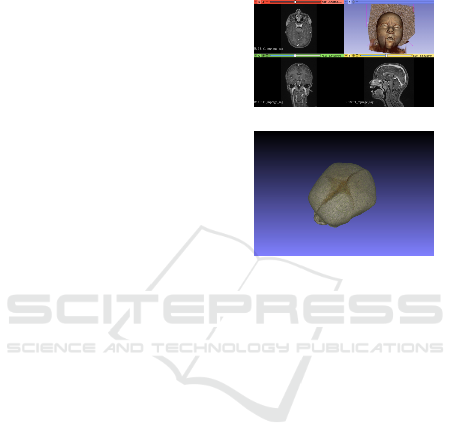
istic geometry of the 9 months old infant obtained at
The University Children Hospital, University of Bel-
grade, Serbia. MRI images consisted of 110 axial MR
slices with 256x256 size and field of view of 240 mm.
The segmentation and meshing was done using soft-
ware packages Slicer and Meshlabs that were then im-
ported as STL files in COMSOL finite-element solver.
Since our main goal is to investigate the effect of
fontanel on the accuracy of inverse model we propose
to use simplified forward model i.e. scalp EEG gen-
erator. In (Gargiulo and Belfiore, 2015) the authors
utilized large scale computational model using large
number of dipoles to simulate EEG signal measured
on the scalp. The inverse models have to rely on much
Figure 1: MRI of Infant Head.
Figure 2: Fontanel Structure.
smaller number of parameters due to the fact the num-
ber of EEG electrodes that can be placed on infant
heads is limited due to the small area. Furthermore,
EEG signals from spatially close electrodes is known
to be highly correlated and thus of limited use in in-
verse EEG models.
To this purpose we propose to use the following
model: a) the regular brain activity is modelled us-
ing 256 Gaussian dipoles placed in the cortical layer
that represent background noise in the EEG signal
measured on the scalp distributed in the circular pat-
tern under the centre of fontanel and b) single cur-
rent dipole model with high power current dipole de-
scribed by three parameters (J
x
,J
y
,J
z
).
Using the finite-element solver in we calculated
the corresponding EM field so that the measurement
model is then given by
y
i j
=
⃗
f (⃗r
i
,t
j
,
⃗
θ) + e
i j
(1)
where y
i j
represents electric potential on the scalp
measured on the ith EEG sensor at timet
j
, f repre-
sents the solution of FE solver at location⃗r
i
and time
t
j
and e
i j
represents the measurements noise/residual
model error. The parameters of the model
⃗
θ are de-
fined by dipole moment and location that are treated
as unknown parameters. As a preliminary approach
we assume that the measurement noise is zero-mean
Gaussian and spatiotemporally uncorrelated. We then
estimate the unknown parameters using maximum
likelihood estimation which in this scenario results
BIOSIGNALS 2024 - 17th International Conference on Bio-inspired Systems and Signal Processing
808
Figure 3: Random instance of dipole moments.
in least-squares estimate by minimizing the error be-
tween measured (simulated and model predicted val-
ues).
⃗
ˆ
θ
LS
= argmin = ∥⃗y −
⃗
f ∥ (2)
where ⃗y is the lumped vector of all the measurements
and
⃗
f is lumped vector of all the model predicted val-
ues. The measurements consist of n spatial measure-
ments and m temporal measurements assuming that
the dipole source is not changing with time.
To evaluate the performance of the proposed al-
gorithms we calculate Cramer-Rao bound which rep-
resents the lower bound on the variance of unbiased
estimators. This bound is a theoretical limit of the
lowest possible variance of the unbiased estimator
and hence it is desirable to have the smallest pos-
sible value as it is a value to which the variance of
the proposed estimator will converge if the number of
measurements is sufficiently high. The Cramer-Rao
bound is calculated using the Fischer information ma-
trix given by
T
i j
(
⃗
θ) = −E
∂lnp
y
(⃗y,
⃗
θ)
∂θ
i
∂lnp
y
(⃗y,
⃗
θ)
∂θ
j
!
(3)
where p(⃗y,
⃗
θ) is the probability density function of the
measurement vector calculated using FEM solver for
a given
⃗
θ. The analytical expression for the Gaus-
sian case can be obtained following (Kay, 1993) using
the Gaussian distribution with a nonlinear parametric
mean.
3 NUMERICAL RESULTS
To simulate the background EEG signal we use cir-
cular grid of 32x32 dipoles with randomly generated
dipole intensities so that in the fontanel region the
background noise dipole density has expected value
of 20µA/cm
2
and standard deviation of 100µA
2
/cm
4
.
In the outer region of the cortex the background EEG
dipoles have expected value of 10µA/cm
2
and stan-
dard deviation of 100µA
2
/cm
4
. For the source we
are trying to estimate/localize we use a dipole with
density of 40µA/cm
2
. Following the values used in
(Gargiulo and Belfiore, 2015) we vary the conductiv-
ity of the fontanel region from 0.01 to 1.51 S/m to
evaluate its effect on the Cramer-Rao bound.
Since in our model the measurement noise is as-
sumed to be Gaussian the Fischer information matrix
and consequently CRLB depend only on the gradient
of the model predicted solution
⃗
f with respect to the
unknown parameters
⃗
θ. We calculate the correspond-
ing gradients using finite difference approximation in
the parameter space. Note that if parameters of in-
terest can be modelled as random variables the cal-
culation of CRLB would require Monte-Carlo simu-
lations. We use the multichannel EEG sensor model
consisting of 4 sensors distributed on the fontanel pe-
riphery. The distance of the source is measured with
respect to the centre of the fontanel structure as illus-
trated in Figure 4.
Figure 4: EEG sensor locations.
1.5
dist
ce
0.5
0.0
1
0
.1
1
c
on
d
u
c
t
i
v
i
ty
Figure 5: Source localization CRB.
In Figure 5 we illustrate the CRB as a function
of distance of the high activity dipole source from
the centre of the fontanel and conductivity. As ex-
pected for larger conductivity values we obtain the
lower CRLB which improves our ability to estimate
the source location accurately. In Figure 6 we illus-
Cramer-Rao Bound for Dipole Source Localization in Infants Using Realistic Geometry
809
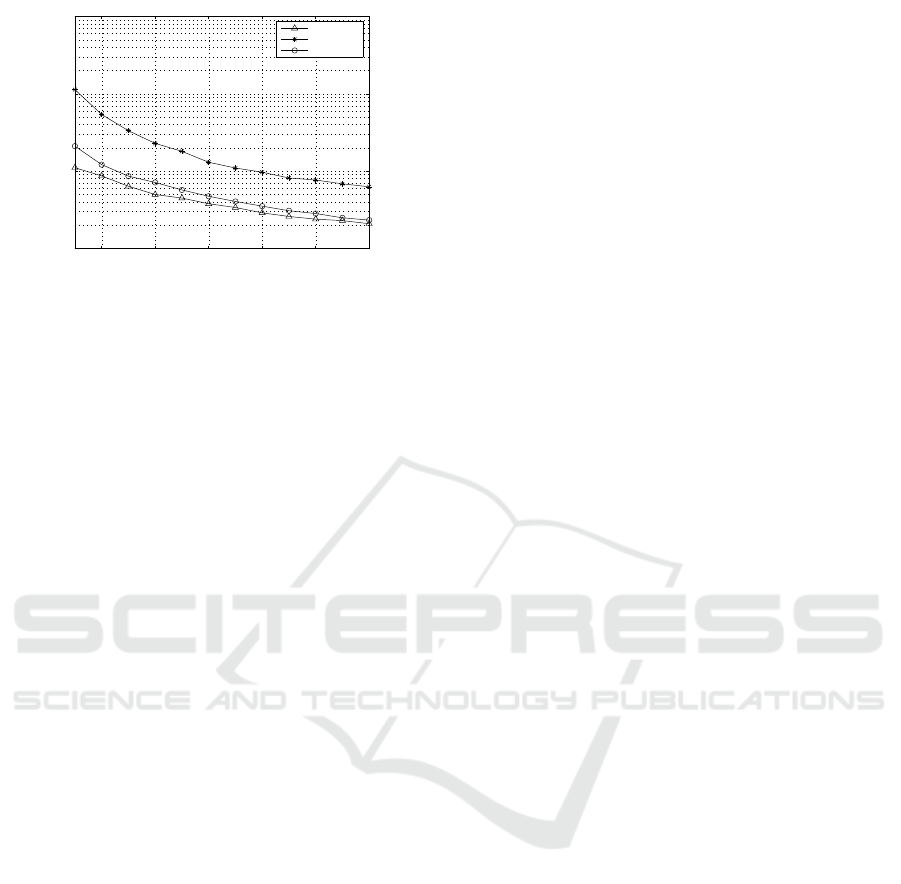
50 100 150 200 250 300
10
−3
10
−2
10
−1
10
0
Number of Measurements
Variances
CRB cond=0.1
LS estimate
CRB Cond=1
Figure 6: Source localization CRB as a function of time.
trate the CRLB and mean square error as a function
of number of measurements. For illustration purposes
we calculate two CRLB for different conductivity val-
ues of 0.1 and 1 and mean square error of LS estima-
tor using 1000 runs. Note that in this example we as-
sume that the dipole intensity is fixed during the mea-
surement interval which may not be valid in realistic
scenario due to the fact that EEG activity epochs are
quite dynamic with respect to time. As expected the
estimator variance decreases with the number of mea-
surements and is expected to asymptotically approach
CRB.
4 CONCLUSIONS
We proposed a computational framework for calculat-
ing theoretical bound for localization of single dipole
source in an infant head using single dipole model
and realistic geometry. The proposed framework en-
ables us to calculate the lowest possible variance that
can be obtained for a given geometry. Our prelim-
inary results indicate that the ability to localize sin-
gle dipole depends on the conductivity as well as ge-
ometry of the subject as well as the conductivity of
fontanel. Therefore an effort should be place on im-
proving our ability to estimate the conductivity jointly
with source localization. In addition, an effort should
be made to investigate the accuracy of the model with
respect to signal-to-noise ratio. Although this analy-
sis is important it is left for future studies as the CRB
of power is significantly dependent on the conductiv-
ity and thus may require improved knowledge on con-
ductivity values of the fontanels. Furthermore, based
on the aforementioned CRB studies the performance
can be significantly increased if we position the EEG
sensors in a such way to minimize CRLB at the largest
possible number of possible source (regions of high
activity) instances. Our results indicate that an ade-
quate localization error can be achieved using inverse
EM modelling approach although it may require ad-
vanced signal processing algorithms.
REFERENCES
Asadzadeh, S., Rezaii, T. Y., Beheshti, S., Delpak, A., and
Meshgini, S. (2020). A systematic review of eeg
source localization techniques and their applications
on diagnosis of brain abnormalitiesl. In J Neurosci
Methods. ELSEVIER.
Cornette, L., Tanner, S., Miall, L., Childs, A., and Arthur,
R. (2002). Magnetic resonance imaging of the infant
brain: anatomical characteristics and clinical signif-
icance of punctate lesions. In Arch Dis Child Fetal
Neonatal. ELSEVIER.
Gargiulo, P. and Belfiore, P. (2015). The effect of fontanel
on scalp eeg potentials in the neonate. In Clin Neuro-
physiol. ELSEVIER.
Gulrajani, R. (1998). Bioelectriciy and Biomagnetism. John
Wiley.
Kay, S. (1993). Fundamentals of Statistical SIgnal Process-
ing. Pearson, New York, 2nd edition.
Malmuvio, J. (1995). Bioelectromagnetism. Oxford Uni-
versity Press.
Shellhaas, R. A. and Clancy, R. R. (2007). Characteri-
zation of neonatal seizures by conventional eeg and
single-channel eeg. In Clinical Neurophysiology. EL-
SEVIER.
Volpe, J. (2001). The Neurology of the Newborn. WB Saun-
ders Co, London, 2nd edition.
BIOSIGNALS 2024 - 17th International Conference on Bio-inspired Systems and Signal Processing
810
