Brain Stroke Prediction Using Visual Geometry Group Model
V. V. L. Narayanan
1
, A. Reddy
1
, V. Venkatesh
1
, S. Tutun
2
, P. Norouzzadeh
1
, E. Snir
2
, S. Mahmoud
3
and B. Rahmani
1,*
1
Saint Louis University, Computer Science Department, St. Louis, MO, U.S.A.
2
Washington University in Saint Louis, Olin Business School, St. Louis, MO, U.S.A.
3
Saint Louis University, Medical School, St. Louis, MO, U.S.A.
Abstract: Stroke has become the leading cause of high mortality and disability rates in the modern era. Early detection
and prediction of stroke can significantly improve patient outcomes. In this study, we propose a deep learning
approach using the Visual Geometry Group (VGG-16) model. VGG-16 is a type of Convolutional Neural
Network (CNN) which is one of the best computer vision models to date to predict the occurrence of a stroke
in the brain. VGG-16 is a type of CNN that is one of the best computer vision models to date. We used a
dataset consisting of Magnetic resonance imaging (MRI) images of patients with and without stroke. The
VGG-16 model was pre-trained on the ImageNet dataset and fine-tuned on our dataset to predict the
occurrence of a stroke. Our experimental results demonstrated that the proposed approach achieves high
accuracy and can effectively predict stroke occurrence. We have also conducted an extensive analysis of the
model's performance and provided insights into important features used by the model to predict stroke
occurrence. The proposed approach has the potential to be used in clinical settings to aid in the early detection
and prevention of stroke.
1 INTRODUCTION
Stroke is a devastating illness that affects millions of
people around the world. According to recent
estimates, over 15 million individuals suffer from this
condition every year. While it is true that stroke is a
major health concern in the United States, with one
person experiencing it every four minutes, this is also
a worldwide problem. Stroke is a leading cause of
death and disability on a global scale, with about 6
million individuals dying from it and another 5
million being left permanently disabled. Clearly,
more needs to be done to prevent and treat this
debilitating illness (WSO, 2022).
A medical emergency commonly referred to as a
brain stroke, brain attack, or cerebrovascular accident
(CVA), occurs when the blood supply to the brain is
interrupted. This can be caused by a blockage or
rupture of a blood artery in the brain. When blood
flow is compromised, brain cells start to die due to a
lack of oxygen and nutrients. This can result in
irreversible brain damage, disability, or even death.
There are two primary subtypes of stroke:
hemorrhagic stroke and ischemic stroke. A
hemorrhagic stroke occurs when a blood vessel in the
*
Corresponding Author
brain bursts and causes bleeding, while an ischemic
stroke occurs when a blood clot blocks an artery in
the brain (A. Kumar, et al., 2023).
High blood pressure, smoking, diabetes, high
cholesterol, obesity, and a family history of stroke or
heart disease are some variables that can increase the
risk of stroke. Lifestyle choices such as poor diet,
inactivity, and stress are also stroke risk factors.
Public education and awareness initiatives can help
reduce the incidence of stroke by promoting healthy
lifestyle choices and encouraging individuals to seek
early medical assistance if they experience stroke
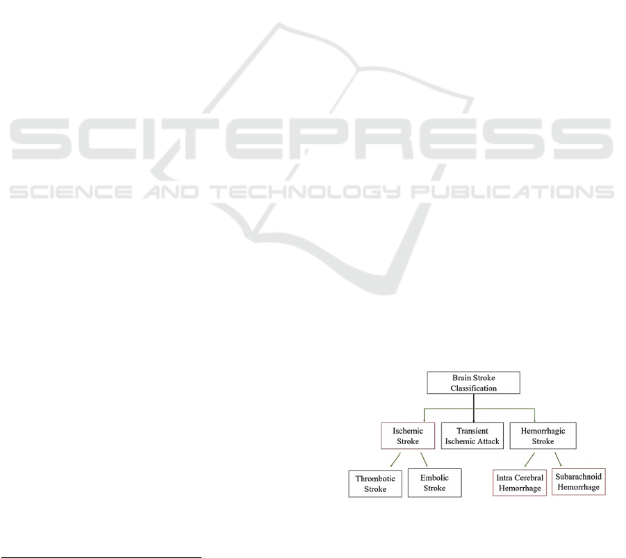
symptoms (R. R. Bailey, 2016). Figure 1 shows the
different type of brain strokes.
Figure 1: Brain Stroke Types – The diagram above
addresses the different types of strokes that can affect a
human brain.
Narayanan, V., Reddy, A., Venkatesh, V., Tutun, S., Norouzzadeh, P., Snir, E., Mahmoud, S. and Rahmani, B.
Brain Stroke Prediction Using Visual Geometry Group Model.
DOI: 10.5220/0012567800003756
Paper published under CC license (CC BY-NC-ND 4.0)
In Proceedings of the 13th International Conference on Data Science, Technology and Applications (DATA 2024), pages 205-210
ISBN: 978-989-758-707-8; ISSN: 2184-285X
Proceedings Copyright © 2024 by SCITEPRESS – Science and Technology Publications, Lda.
205

Artificial intelligence (AI) has advanced
significantly in recent years, with applications in
everything from autonomous vehicles and medical
diagnostics to voice and image recognition. The
advancement of deep learning, a branch of machine
learning that has made strides in a variety of AI
applications, has been a major force behind this
development. We go over the fundamentals of deep
learning, its uses, and its possible social effects in this
paper.
Deep learning is a vital component of Artificial
Intelligence, which uses artificial neural networks to
model and solve complex classification problems.
These artificial neurons, which function as the
processing and transformation units of these neural
networks, are organized into numerous layers of
interconnected nodes. Deeper layers of neurons learn
to recognize increasingly abstract and complicated
patterns as they learn to recognize and extract
different characteristics of the data.
Deep Learning has the capacity to learn and
improve on its own, without being explicitly
programmed. By processing large amounts of data
and identifying patterns and relationships within that
data, deep learning algorithms can learn to perform
complex tasks such as image and speech recognition,
natural language processing, and even game playing.
In healthcare, deep learning is being used to
develop diagnostic tools to analyse medical images
and data to detect diseases such as cancer and
Alzheimer’s (I. M. Sheikh and M. A. Chachoo, 2022).
These tools have the potential to improve the
accuracy and speed of diagnosis, enabling earlier
detection and better outcomes for patients (L Chen, et
al., 2023; P Bentley, et al., 2014).
Yoon-A Choi et al. proposed a system for
predicting the likelihood of stroke based on real-time
bio-signal data using neural networks. 3,322
electroencephalogram (EEG) and
(electrocardiogram) ECG signals were collected from
stroke patients and healthy individuals. The proposed
model is a Convolutional Neural Network (CNN),
which extracts features from the signals and a long
short-term memory (LSTM) network that models
temporal dependencies. This system gave a model
accuracy of - 93.9 percent and a sensitivity of 96.7
percent (Y-A Choi, et al., 2021). This research
however needed further research to validate the
system’s effectiveness in larger and more diverse
populations.
Vivek S Yedavalli et al. discussed the potential
applications of artificial intelligence (AI) in stroke
imaging, including diagnosis, treatment selection,
and prognosis prediction, using different machine
learning models and neural networks. Models like
Convolutional Neural Networks (CNN), Long Short-
Term Memory (LSTM), Recurrent Neural Networks
(RNN), Support Vector Machines (SVM), and
Random Forest (RF) were trained, and their
accuracies were compared to select the best model. It
was shown that CNN had the highest accuracy at 91
percent. This study used 4 different datasets, namely,
MRI-GENIE, STRIDE, MR CLEAN, and TRACK-
TBI (B B Ozkara, et al., 2023).
Hilbert et al. proposed a deep learning model for
predicting the outcome of endovascular treatment in
patients with acute ischemic stroke. The dataset was
a collection of 92 patients who underwent
endovascular treatment for acute ischemic stroke and
were divided into a training set of 60 patients and a
test set of 32 patients. To improve its performance on
the outcome prediction task, the deep learning model
was trained on a small subset of the training set
(n=10) utilizing transfer learning and fine-tuning
approaches. The model was then assessed using the
test set and the remaining training data. This proposed
system used the VGG-16 model. The model was then
fine-tuned on the training data using a transfer
learning approach, which involves using the pre-
trained weights of the VGG-16 model and training the
final layers on the task-specific dataset. This gave an
accuracy of 80 percent (A Hilbert, et al., 2019).
Sonavane et al. presented a method for detecting
brain stroke using convolutional neural networks
(CNNs) and deep learning models. This is a novel
approach that combines CNNs with deep learning
models to automatically see brain strokes from
computed tomography (CT) images. The dataset is a
collection of 250 CT images, including 150 healthy
and 100 stroke images, which were used to train and
evaluate their deep-learning models. The
methodology tested four different deep learning
models, including a CNN, a deep belief network, a
stacked autoencoder, and a convolutional
autoencoder, to determine the most effective model
for detecting brain stroke. The CNN model achieved
the highest accuracy at 97.6 percent for detecting
brain stroke, followed by the stacked autoencoder
model with an accuracy of 94.8 percent. The authors
also performed a comparative analysis with existing
methods and found that their proposed method
outperformed existing methods for brain stroke
detection. This proposed system’s future research
could explore the use of larger datasets and more
advanced deep learning models to further improve the
performance of the brain stroke detection system (B
R Gaidhani, et al., 2019).
DATA 2024 - 13th International Conference on Data Science, Technology and Applications
206

Mahadevan et al. compared the performance of
traditional hand-crafted features and convolutional
neural networks (CNNs) for diagnosing stroke from
retinal images. The dataset that was used in this
proposed system contained 450 retinal images,
including 150 healthy, 150 hypertensive, and 150
diabetic images, to train and evaluate their models.
The methodology involved in this method comprised
of testing two different approaches: traditional feature
extraction using hand-crafted features, and deep
learning using a CNN. The results of the study
showed that the CNN achieved significantly better
performance than the traditional feature extraction
method, with an accuracy of 96.5 percent compared
to 87.3 percent for the hand-crafted features. The
authors also compared the two approaches and found
that the CNN had higher sensitivity, specificity, and
F1 score for diagnosing stroke from retinal images (R
S Jeena, et al., 2021).
Amitava Nag and his research group did another
research to predict the brain stroke. They applied
Ada-Boost and other boosting methods to investigate
and classify the information of more than 48000
patients achieving high accuracy (S. Mondal, et al.,
2023). The main difference between the current and
Nag’s project is the type of data. We worked on image
data.
In the other similar project, Sachin and Vishal
Jain classified brain tumours with deep learning
models with 98% accuracy. The applied 5, 10 and 20
cross validation folds to realize high accuracy (S. Jain
and V. Jain, 2023). Our project predicts the brain
stroke with Neural Network models.
Emotion recognition project using VGG-16
model accomplished by Srindhar and his research
group in 2023 (S. Vignesh, et al., 2023). Sunil Kumar
et al, applied random forest and VGG-16 methods to
classify bell pepper leaf disease with LBP features
(M. Bhagat, et al., 2023).
Other researchers applied VGG-16 method to
classify and predict different types of diseases and
syndromes, with different type of dataset, and
accuracy. What makes our project different is that we
predict brain stroke using image data with high
accuracy. For this aim we applied different methods,
which VGG-16 predicted the stroke with highest
accuracy among other methods. We worked on a
large image dataset.
*
https://www.kaggle.com/datasets/afridirahman/brain-
stroke-ct-image-dataset
2 DATA DESCRIPTION
The dataset used for this project is collected from
Kaggle
*
, an online community, with millions of
diverse datasets available for analyses. We chose a
collection of medical images, specifically computed
tomography (CT) images, of the brain of individuals
who have experienced a stroke and of individuals
who have not experienced a stroke. There are a total
of 2,501 images in the datasets out of which 1,551
belong to individuals who have not experienced
strokes and the remaining 950 belong to individuals
who have experienced a stroke. This is a binary
classification problem where images belong to two
different classes.
3 METHODOLOGY
3.1 Data Pre-Processing
The final image size of the input dataset for the VGG-
16 (Visual Geometry Group) model is 256x256
pixels. In the next pre-processing step we split our
dataset into training (80 percent), testing (10 percent),
and validation (10 percent) sets. To include more
samples in our dataset we implemented an image data
generator that creates different variations of each
image at each epoch. The variations include random
image rotations, horizontal flips, and shifts.
Additionally, zoom and brightness effects are set in
the range of 0.2 and 0.8. After the pre-processing
steps, the transformed images are used by the VGG-
16 model to predict stroke.
3.2 Baseline Model and Methodology
The VGG-16 model which is a 16-layered deep
image classification convolutional neural network
(CNN) architecture is the baseline model in our
research. It is one specific variation of a
Convolutional Neural Networks (CNN), originally
proposed by Simonyan and Zisserman (K Simonyan
and A Zisserman, 2019). This CNN was chosen in
this study based on its prior success at image
recognition. While many variations exist for CNN,
using a configuration that has been previously
validated enables achieving optimal results. The
VGG-16 model differs from other configuration in
its choice of convolutional layers, pooling layers,
and dense layers. In all, VGG-16, has 16
Brain Stroke Prediction Using Visual Geometry Group Model
207
convolutional layers. These are the layers where
learning occurs. This specification facilitates up to
138 parameters that can be trained. The 16
convolutional layers are divided in 5 groups, each
with a pooling layer at the end. At the end of the
stack of layers there a three dense layers. In all,
VGG-16 has 21 layers. In addition to the predefined
layers in the CNN, VGG-16, the convolution layers
are relatively small, with 3x3 filters with a stride
of 1.
The pre-trained VGG-16 model classifies over
1000 images from different categories. We
incorporated the VGG-16 model into our sequential
model with several flattened, dense and output layers.
The output layer consists of a sigmoid activation
function, which is used for binary classification that
consists of two classes i.e. stroke and non-stroke.
Model training was done on 25 epochs and a batch
size of 32. The final sequential model used a
monitoring metric called early stopping that halts the
training process when there is no further
improvement in learning.
In this proposed system, we use ‘Adam’ optimizer
for our model because the Adam optimizer
dynamically adjusts for each parameter based on the
first and second moments of the gradients, which
increases the efficiency of the model performance and
simultaneously, requires low storage space. We
calculate the loss by the binary cross-entropy metric,
which computes gradients correctly and encourages
classification with high accuracy rate.
The model also makes use of early stopping,
which creates an early stopping call back that
monitors the validation accuracy and stops the
training process if the accuracy does not improve for
a specified number of epochs.
4 RESULTS
During the first epoch, the training accuracy started at
61 percent which increased significantly to 83 percent
at the end of the 25th epoch. While predicting
outcomes on the test dataset, the model was able to
correctly predict over 80 percent of the outcomes.
Since the difference between the training and test
accuracies is not too high, we can say that the model
is not prone to overfitting.
From the classification report, the precision for
people with no brain stroke is 0.90, and for the people
with brain stroke is 0.77. This means that there are
more people not detected with brain stroke and fewer
people with brain stroke.
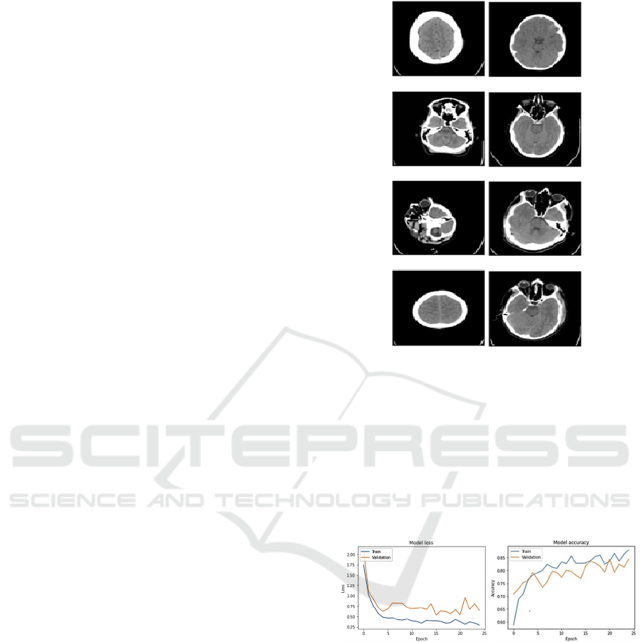
Figure 2: Brain Stroke Detection – The left image
represents the images with no brain stroke and the right-side
image represents images with the prediction of a brain
stroke.
Figure 2 portrays the difference between a normal
brain and a brain that has stroke. The image on the
left side represents the scans of the patients with no
stroke prediction and the images in the right side are
that of the people who have experienced a brain
stroke.
Figure 3: The AUC for Training Vs Validation Loss.
Figure 3 is a graphical representation of the
performance of the Visual Geometry Group Model
during the training and validation phases. This
measures the model’s ability to correctly classify data
points. It is plotted against the number of epochs of
the training process. The image on the left represents
the model loss and exhibits that the model performed
well without any overfitting until the 5
th
epoch, as the
training and validation loss are close enough to each
other. The image on the right represents the model
accuracy, which measures the overall performance of
the model.
DATA 2024 - 13th International Conference on Data Science, Technology and Applications
208
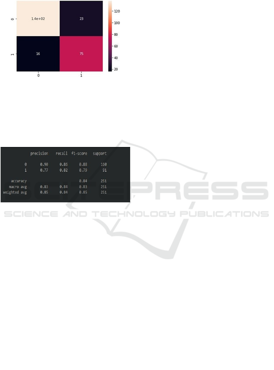
Figure 4: Confusion Matrix.
Figure 4 is a confusion matrix, that compares the
actual and predicted values of the model on the given
dataset. The confusion matrix displays the least
number of true positives. The number of true
negatives is more than the number of true positives,
which means that there are more predictions for
people with brain stroke.
Figure 5: Classification Report.
Figure 5 portrays the classification report and
provides a detailed evaluation of the model’s ability
to correctly classify instances as is typically used in
classification tasks where the goal is to predict the
class of the given dataset. The proportion of the
positive predictions that actually had stroke is 0.90
and the proportion of actual positive instances that
were correctly identified by the model is 0.86.
5 CONCLUSION
In conclusion, the VGG-16 model shows promising
results in predicting the occurrence of brain stroke
using medical imaging data. Our research
demonstrates that the VGG-16 model can achieve
high accuracy of 80 percent in predicting the
likelihood of stroke by analysing the images of the
brain without overfitting. However, further research
is needed to improve the accuracy and
generalizability of the VGG-16 model, as well as to
explore its potential for other medical imaging
applications. Additionally, it is important to address
ethical and privacy concerns related to the use of
patient data in developing and deploying AI models
for medical diagnosis. Overall, our research suggests
that the VGG-16 model has significant potential as a
tool for predicting brain stroke using medical imaging
data and underscores the importance of continued
research and development in this area.
6 FUTURE SCOPE
In this project we applied neural networks to predict
the stroke. In the next project we apply another
prediction and image analysis methods to validate our
results with higher accuracy. There is a plan to
develop a platform to upload images and define the
possibility of brain stroke.
Data Availability
The dataset used for this project is collected from
Kaggle. https://www.kaggle.com/datasets/afridirahm
an/brain-stroke-ct-image-dataset.
Conflict Interest Statement
There is no conflict of interest declared by authors.
All authors have reviewed and agreed with
manuscript. We state that the submission is original
paper and is not under review at any other journal.
Funding
No Funding has been applied for this project.
Ethical Approval
All subjects gave their informed consent for inclusion
before they participated in the study.
Consent to Participate
Authors consent to participate in this project and we
know that: the research may not have direct benefit to
us. Our participation is entirely volunteer. There is a
right to withdraw from the project at any time without
any consequences.
Consent to Publish
We give our consent for the publication of exclusive
details, that could be included figures and tables and
details within the manuscript to be published in
Computational Brain & Behavior.
Brain Stroke Prediction Using Visual Geometry Group Model
209

REFERENCES
Global Stroke Fact Sheet, World Stroke Organization
(WSO), 2022.
A. Kumar, A. Unnithan, J. M Das, Parth Mehta.
Hemorrhagic Stroke, StatPearls, 2023
R. R. Bailey, Lifestyle Modification for Secondary Stroke
Prevention, PubMed, 2016
I. M. Sheikh, M. A. Chachoo, An enforced block diagonal
low-rank representation method for the classification of
medical image patterns, International Journal of
Information Technology, 2022.
L Chen, P Bentley, D Rueckert, Fully automatic acute
ischemic lesion segmentation in DWI using
convolutional neural networks. NeuroImage: Clinical.
Retrieved February 27, 2023
P Bentley, J Ganesalingam, AL Carlton, K Mahady, S
Epton, P Rinne, P Sharma, O Halse, A Mehta, P
Rueckert, Prediction of stroke thrombolysis outcome
using CT brain machine learning. Neuroimage Clin.
2014 Mar 30;4:635-40. doi: 10.1016/j.nicl.2014.
02.003. PMID: 24936414; PMCID: PMC4053635.
Y-A Choi, S-J Park, J-A Jun, C-S Pyo, K-H Cho, H-S Lee,
J-H Yu, Deep Learning-Based Stroke Disease
Prediction System Using Real-Time Bio Signals,
Sensors, 2021
B B Ozkara, M Karabacak, O Hamam , R Wang, A Kotha,
N Khalili, M Hoseinyazdi, M M Chen, M Wintermark,
V S Yedavalli, Prediction of Functional Outcome in
Stroke Patients with Proximal Middle Cerebral Artery
Occlusions Using Machine Learning Models, Pubmed,
2023
A Hilbert, LA Ramos, HJA Van Os, et, Data-efficient deep
learning of radiological image data for outcome
prediction after endovascular treatment of patients with
acute ischemic stroke, Computers in Biology and
Medicine, 2019
B R Gaidhani, R R Rajamenakshi, S Sonavane, Brain
Stroke Detection Using Convolutional Neural Network
and Deep Learning Models, Conference: 2019 2nd
International Conference on Intelligent Communication
and Computational Techniques (ICCT), 2019
R S Jeena, G Shiny, A S Kumar, K Mahadevan, A
Comparative analysis of stroke diagnosis from retinal
images using hand-crafted features and CNN, Journal
of Intelligent and Fuzzy Systems 41(3):1-9, 2021
S. Mondal, S. Ghosh, A. Nag, Brain stroke prediction
model based on boosting and stacking ensemble
approach, International Journal of Information
Technology, 2023.
S. Jain, V. Jain, Novel approach to classify brain tumor
based on transfer learning and deep learning,
International Journal of Information Technology, 15,
pages 2031–2038, 2023.
S. Vignesh, M. Savithadevi, M. Sridevi, R. Sridhar, A novel
facial emotion recognition model using segmentation
VGG-19 architecture, International Journal of
Information Technology volume 15, pages1777–1787
(2023).
M. Bhagat, D. Kumar, S. Kumar, Bell pepper leaf disease
classification with LBP and VGG-16 based fused
features and RF classifier, International Journal of
Information Technology volume 15, pages465–475
(2023)
O Ozaltin, O Coskun, O Yeniay, A Subasi, A Deep
Learning Approach for Detecting Stroke from Brain CT
Images Using OzNet. Bioengineering. 2022; 9(12):783.
K Simonyan, A Zisserman, Very deep convolutional
networks for large-scale image recognition. arXiv
preprint arXiv:1409.1556. 2019
DATA 2024 - 13th International Conference on Data Science, Technology and Applications
210
