
Modeling of an Electric-Based Defogging System for Laparoscopy
Ali Al Hadi Orabi Al Haddad
1
, Nour Mansour
1
, Wassim Salameh
2
, Ali Cherry
3
, Bassam Hussein
4
,
Houssein Hajj Hassan
5
and Mohamad Hajj-Hassan
1
1
Department of Biomedical Engineering, Lebanese International University, Bekaa, Lebanon
2
Department of Mechanical Engineering, The International University of Beirut, Beirut, Lebanon
3
Department of Biomedical Engineering, The International University of Beirut, Beirut, Lebanon
4
Department of Industrial Engineering, The International University of Beirut, Beirut, Lebanon
5
Faculty of Health Sciences, Al Maaref University, Beirut, Lebanon
Keywords: Laparoscopy, Defogging, Modeling.
Abstract: This paper addresses the common issue of laparoscopic lens fogging (LLF), which hampers the surgeon's
visibility and prolongs procedures. The proposed solution involves an advanced electrical defogging system
utilizing a thin, conductive transparent material in different configurations such as circular disk and concentric
spiral models. These configurations generate consistent heat on the lens surface, minimizing the temperature
difference between the lens and the inside of the body. The ideal results were achieved with a 14-circle ITO
model, maintaining a constant 310 K (37°C) on the lens surface. Through detailed simulations and modeling,
this research demonstrates the effectiveness of the proposed system in maintaining a fog-free lens, ultimately
reducing operational delays and enhancing both surgical efficiency and patient safety in laparoscopic
procedures.
1 INTRODUCTION
Laparoscopy is a minimally invasive surgical
technique that relies heavily on the clarity of the
laparoscopic lens to provide surgeons with a clear
view of the operative field. It is conducted in response
to view internal organs of the body clearly such as
checking for polyps in stomach, bleeding, infections,
and blockages. The main advantages of this surgery
are faster recovery times and less bleeding compared
to exploratory laparotomy leading to shorter stay at
the hospital. Fogging of the laparoscope lens is a
prevalent issue in laparoscopic surgery. It arises from
the temperature disparity between the human body
and the laparoscope lens, leading to the condensation
of water vapor on the cold lens leading to obstructing
the view. Multiple methods exist that solve this issue
but none has the efficiency to provide both a totally
clear image and full safety of the patients with zero
side effects. Several anti-fogging approaches have
been established and are used in the laparoscopic
surgical field. Physical methods include preheating
the endoscope camera with 60-80℃ sterile normal
saline for 5-10 minutes before insertion. Chemical
methods involve applying substances like iodine,
anti-fog oil, and alcohol to the lens. Ventilation
systems work by maintaining airflow to prevent
fogging using CO2 (Calhoun & Redan, 2014). Each
method has its advantages and drawbacks. Physical
methods can be time-consuming and lack
effectiveness but are the cheapest among all methods
(Yasui & Kubo, 2022), while chemical methods may
leave residues that harm the lens or cause irritation to
the patient but can have a very clear image (World
Laparoscopy Hospital, 2013). Ventilation systems
can cause heart and other health issues to the patient
but also provides a clear image (Jiang & Sun, 2019).
The aim of this research is to investigate defogging
using heating to provide a clearer and more visible
image during laparoscopic surgeries with minimal
side-effects.
2 METHODOLOGY
The proposed defogging system employs a thin
conductive Indium Tin Oxide (ITO) layer to generate
heat over the laparoscopic lens, maintaining a
constant temperature of 310 K (37°C), which is safe
for both human cells and the laparoscopic lens. This
Al Haddad, A. H. O., Mansour, N., Salameh, W., Cherry, A., Hussein, B., Hassan, H. H. and Hajj-Hassan, M.
Modeling of an Electric-Based Defogging System for Laparoscopy.
DOI: 10.5220/0013133200003911
Paper published under CC license (CC BY-NC-ND 4.0)
In Proceedings of the 18th International Joint Conference on Biomedical Engineering Systems and Technologies (BIOSTEC 2025) - Volume 1, pages 139-142
ISBN: 978-989-758-731-3; ISSN: 2184-4305
Proceedings Copyright © 2025 by SCITEPRESS – Science and Technology Publications, Lda.
139

system prevents fogging by reducing the temperature
difference between the body and the lens to near zero,
effectively eliminating condensation. The research
approach includes defining boundary conditions,
implementing electrical and heat transfer models, and
conducting simulations to optimize the ITO
configuration. COMSOL Multiphysics software is
used for modeling the electric-based defogging
system. Detailed simulations are conducted to model
the temperature distribution and effectiveness of the
system, considering various parameters such as film
thickness, electrical properties, and heating
efficiency. The modeling starts with sketching a 10
mm diameter circle extruded by 1mm as thickness
which will represent the lens. Followed by sketching
another circle over the lens with approximately 10
mm diameter and later replaced by concentric spiral
circles with 10 μm thickness for both which will be
the ITO layer. The lens is made of silica glass which
withstands up to 1100°C and has many other
characteristics that make it suitable for this project.
And as for the ITO it was chosen in this project due
to its high transparency which peaks around 93% and
always above 80% throughout all the visible spectrum
as shown in figure 1. while having high electrical and
thermal conductivity to be able to generate heat using
current by utilizing joule’s effect. After everything is
in place we begin to assign the electrical properties
initializing with having an electric potential of 5V as
a constant input which is used in laparoscopy
machines and assigning a ground. Each is assigned
over 1 of the edges of the ITO circuit as shown in
Figure 2 note that both edges indicated by the arrows
should be connected to external pads using a wire
bonding machine. Additionally, for the circular disk
model, two pads should be placed directly on the
opposite edges of the disk for voltage and ground
interfacing and connections. DC current is used in this
simulation which generates more heat than AC
current. Heat transfer in solids is used to simulate the
heat transfer between the ITO and the lens to finally
Figure 1: Transmission spectra of a typical ITO thin film
(Kim, 2023).
Figure 2: Assignment of electric potential and ground on
ITO spiral.
get the results. Design optimization is applied to the
concentric circles by increasing the number of circles
placed on the lens to get the ideal result which is full
distribution of 310K over all the lens which is the
human body temperature.
3 RESULTS AND DISCUSSION
The simulations of the first circular configuration will
be observed, then using the second spiral
configuration numerous results have been derived
while optimizing the size of ITO in order to detect
variable results reaching the ideal outcome.
Figure
3
: Temperature distribution on the ITO circular full
disc.
After observing the above simulation in Figure 3, it is
noticeable that the temperature is disturbed in a very
inadequate and inefficient manner reaching a max
temperature of 307 K only on the center of the lens.
Thus, circular disc configuration that is used is no
longer applicable to reach the most ideal and desired
results needed for this research.
DC Voltage
BIODEVICES 2025 - 18th International Conference on Biomedical Electronics and Devices
140
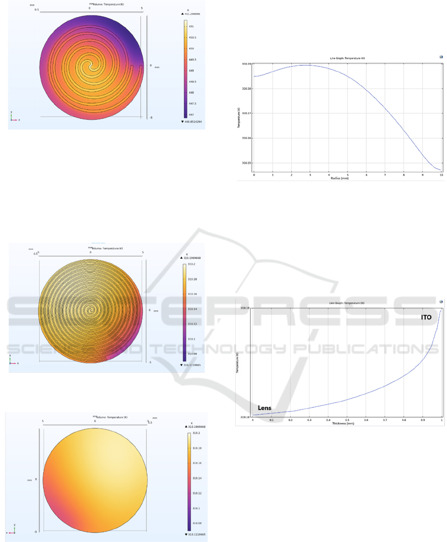
Figure
4
: Temperature distribution on the 3 circles ITO
spiral.
Figure 4 shows the temperature distribution on the
three circle ITO spiral and on the laparoscopic lens
respectively. It can be noticed that a high temperature
of 451 K is reached only on the middle of the lens and
gradually decreasing reaching a minimal value of 447
K on the edges.
Figure
5
: Temperature distribution on the 14 circles ITO
spiral.
Figure
6
: Temperature distribution on the lens of 14 circles
ITO model.
Figures 5 and 6 show the ideal temperature
distribution on the 14 circles ITO spiral and on the
laparoscopic lens respectively. Observing that a
desired maximal temperature of 310.2 K is distributed
on the whole lens, bear in mind that there is a
negligible difference of 0.08 K on the lower edge
reaching a value of 310.14 K.
Figure
7
: Line graph representing temperature distribution
with respect to the radius of the lens.
Figure 7 shows the temperature distribution on the
laparoscopic lens in Kelvin while using 14 circles
ITO model with respect to the radius of the lens in
mm. Noting that it reaches a peak of 310.19 K at a 3
mm radius near to the center of the lens.
Figure
8
: Line graph representing temperature distribution
with respect to the thickness of the lens.
Figure 8 shows the temperature distribution on the
laparoscopic lens in Kelvin while using 14 circles
ITO model with respect to the thickness of the lens in
mm. It demonstrates that the temperature is higher on
the ITO side, as the heat generated by the ITO, which
is embedded within the laparoscope, gradually
transfers to the lens.
The first simulation with a circular ITO disc
resulted a deficient and inadequate temperature
distribution on the lens with a temperature of 307 K.
The second configuration that have a spiral form,
multiple simulations have been resulted initializing
with 3 circles ITO then increasing the number of
Modeling of an Electric-Based Defogging System for Laparoscopy
141

circles to attempt 5, 8, 10 and ending with 14 circles
of ITO. At first, 3 circles ITO have shown an
insufficient temperature distribution with an
extremely high temperature of 451 K. As the number
of circles increases, the temperature distribution
becomes better and more intensified because less
distance is found between turns. In addition, the
temperature value is decreasing due to increasing the
length of the wire. This can be noticed by comparing
the 3-circle configuration with the 14 circles, where
the distribution being uniform all over the surface and
the temperature dropped from around 450K to 310K.
The 14 circles ITO configuration has the ideal
temperature reached with a value of 310.2 K
representing body temperature that is perfectly and
equally distributed on the whole lens where such
result is emitted by applying an input voltage of 5 V
only. Thus, the 14 circles configuration was the
optimized result. In addition, it is important to
mention that as the thickness of the laparoscopic lens
increases moving toward the ITO, the temperature
distribution increases as shown in figure 8 and vice
versa moving away from the ITO spiral thus the
temperature distribution will keep decreasing till
reaching the lens. Finally, after the above analysis and
interpretation of the whole results and after reaching
our optimal distribution. It is essential to highlight on
the main points that specializes our research from any
other available techniques that improve vision during
laparoscopic surgeries. This research will initially add
a special advantage of keeping a clear and vivid
image during the whole laparoscopic surgical
procedure. Adding, it will maintain a safe and secured
heating technique on the lens since it is found on the
inside of the laparoscope keeping the patient well and
protected without any side effects compared to any
other technique.
4 CONCLUSIONS
The development of an electric-based defogging
system for laparoscopy represents a significant
advancement in surgical technology. This research
demonstrates that the proposed system can effectively
maintain a clear lens surface, addressing a common
issue that hinders laparoscopic surgeries while not
harming the patient with any side effects. Future work
should focus on the testing of the system under real-
life conditions to validate its performance and
reliability. Further optimization and experimental
validation are necessary to enhance system
performance and ensure practical applicability.
ACKNOWLEDGEMENTS
We would like to acknowledge the financial support
from the XNY Medical, a manufacturer and
distributor of minimal invasive surgery (MIS)
medical devices, China.
REFERENCES
Calhoun, J., and Redan, J. (2014, January). Elimination of
Laparoscopic Lens Fogging Using Directional Flow of
CO2. Journal of the society of laparoscopic and
Robotic Surgeons, 18. Retrieved from https://www.
ncbi.nlm.nih.gov/pmc/articles/PMC3939343/
Yasui, T., and Kubo, Y. (2022). Mechanism underlying lens
fogging and its countermeasure in laparoscopic surgery.
Minimally Invasive Therapy and Allied Technologies,
32(2), 56-61.
World Laparoscopy Hospital. (2013, February 3).
Retrieved from https://www.laparoscopyhospital.com/
news/preview.php?id=93#:~:text=Ventilation%20Syst
ems%20-%20Ventilation%20systems%20are,the%20l
ens%20and%20creating%20fog
Jiang, R., and Sun, Y. (2019). Effect of different carbon
dioxide (CO2) insufflation for laparoscopic colorectal
surgery in elderly patients. National Library Of
Medicine, 98(41).
Kim, M.-J. (2023). A Study on Optimal Indium Tin Oxide
Thickness as Transparent Conductive Electrodes for
Near-Ultraviolet Light-Emitting Diodes. MDPI,
16(13), 4718.
BIODEVICES 2025 - 18th International Conference on Biomedical Electronics and Devices
142
