
Pneumonia Detection in X-Ray Chest Images Based on Convolutional
Neural Networks and Data Augmentation Methods
Samia Dardouri
1,2 a
1
Department of Computer Science, College of Computing and Information Technology, Shaqra University, Saudi Arabia
2
InnoV'COM Laboratory-Sup'Com, University of Carthage, Tunisia
Keywords: CNN, Feature Extraction, Pneumonia Infection, Image Data Augmentation, Deep Learning, Adam Optimizer,
Early Detection.
Abstract: Pneumonia, a widespread lung ailment, stands as a leading global cause of mortality, particularly affecting
vulnerable demographics such as children under five, the elderly, and individuals with underlying health
conditions. Accounting for a significant portion of childhood fatalities, at 18%, pneumonia remains a critical
health concern. Despite advancements in imaging diagnostic methods, chest radiographs remain pivotal due
to their cost-effectiveness and rapid results. The proposed model, trained on data sourced from a readily
available Kaggle database, consists of two primary stages: image preprocessing and feature extraction/image
classification. Utilizing a CNN model, the framework achieves remarkable performance metrics, with
precision, recall, F1-score, and accuracy reaching 93%, 96%, 94%, and 96%, respectively. These results
underscore the CNN model's effectiveness in pneumonia detection, showcasing superior consistency and
accuracy compared to other pretrained deep learning models.
1 INTRODUCTION
Pneumonia is a prevalent and potentially life-
threatening respiratory infection, particularly
affecting vulnerable populations such as children, the
elderly, and immunocompromised individuals. Early
detection of pneumonia is crucial for timely
intervention and treatment to prevent complications
and improve patient outcomes. While numerous
imaging diagnostic methods exist, many are
prohibitively expensive and inaccessible to large
segments of the population, especially in low-
resource regions Asnake, N.W (2024). Additionally,
the shortage of radiology experts in these areas and
long waiting times for diagnoses exacerbate the
severity of the disease and contribute to increased
mortality rates. Diagnostic radiography, although
cost-effective and rapid, may lead to
misinterpretations due to visualized opacities.
To address these challenges, recent studies have
explored machine learning techniques, particularly
deep learning models like convolutional neural
networks (CNNs), to aid in pneumonia diagnosis
using high-resolution imaging modalities such as
a
https://orcid.org/ 0000-0002-1376-9607
computed tomography (CT) scans. One such study
developed a deep learning architecture tailored for
diagnosing severe pneumonia cases from chest X-
rays. Leveraging a dataset from the Radiological
Society of North America, this study focused on
specialized zones within the chest X-rays for
improved diagnostic accuracy.
In recent years, the healthcare landscape has
witnessed the emergence of various technologies
such as genomics and imaging, which have brought
forth vast and intricate datasets in Asnake, N.W.,
Salau (2024). While chest X-ray images remain a
primary diagnostic tool for pneumonia, they can pose
challenges due to their nuanced nature, sometimes
leading to misclassifications by expert radiologists
and subsequent incorrect treatments (Lamia A, Fawaz
A (2022)). This underscores the need for an automatic
and intelligent model to aid radiologists in accurately
diagnosing different types of pneumonia from chest
X-ray images Goyal, S., Singh, R (2023).
Deep learning, a subset of machine learning
inspired by the brain's structure and function, has
emerged as a powerful tool in medical image analysis
Kareem, A., Liu, H (2022). These algorithms excel at
Dardouri, S.
Pneumonia Detection in X-Ray Chest Images Based on Convolutional Neural Networks and Data Augmentation Methods.
DOI: 10.5220/0013147300003938
In Proceedings of the 11th International Conference on Information and Communication Technologies for Ageing Well and e-Health (ICT4AWE 2025), pages 165-172
ISBN: 978-989-758-743-6; ISSN: 2184-4984
Copyright © 2025 by Paper published under CC license (CC BY-NC-ND 4.0)
165

quantifying, identifying, and classifying patterns
within medical images by learning features directly
from data, eliminating the need for manual feature
design based on domain-specific knowledge.
Convolutional neural networks (CNNs) are a
prominent example of deep learning models utilized
in this context. These layers specialize in processing
images and extracting low-level features, such as
edges, while efficiently capturing temporal and
spatial dependencies with the aid of filters. Unlike
traditional feed-forward layers, CNNs significantly
reduce computational complexity by sharing weights
and utilizing fewer parameters. As a result, CNNs
offer an effective approach for medical practitioners
to diagnose and classify specific medical conditions
with accuracy (Z. Li et al 2019 ; M. K. Gourisaria
2023). The structure of the paper is as follows:
Section 2 provides a detailed review of related works,
summarizing relevant research on the topic. Section 3
describes the dataset utilized in this study. Section 4
outlines the proposed methodology implemented in
the research. Section 5 examines the results and
compares them with findings from recent studies.
Finally, Section 6 concludes the paper by
summarizing key insights and proposing directions
for future research.
2 RELATED WORKS
In the field of disease detection, numerous
researchers have been actively engaged in developing
automated detection models. Deep learning
techniques have emerged as valuable tools for
enhancing productivity, especially in computer-
assisted diagnosis technologies, notably within
medical imaging, image classification, and image
restoration Venkateswara Reddy. (2022). Author in
Shagun Sharma. (2023) proposed deep learning (DL)
model comprises several stages: data collection,
preprocessing, feature extraction, training, testing,
classification, and pneumonia prediction. During data
preprocessing, the data is balanced and normalized,
ensuring it falls within a normalized range of [0-255].
Subsequently, the normalized data is inputted into the
VGG16 model for feature extraction Liu, Y (2023).
This step involves extracting pertinent features from
the images, facilitating the classification and
prediction process. With its 16 layers encompassing
input, convolution, pooling, dense, and output layers,
VGG16 enables comprehensive feature extraction.
The significant challenges faced in pneumonia
detection include the large number of patients and the
shortage of medical experts and supporting staff. The
development of deep learning-based methods for
early detection of pneumonia has garnered significant
attention in recent years due to their potential to
improve diagnostic accuracy and efficiency. By
leveraging advanced computational techniques and
large datasets of annotated medical images,
researchers have made significant strides in
developing deep learning models capable of detecting
pneumonia infections at an early stage Shadi A.
(2022).
In the study discussed in Lamia A. (2022),
pneumonia emerges as a rapidly spreading disease,
posing significant risks to individuals' health and
well-being. Biomedical diagnosis of pneumonia
typically involves a range of diagnostic tools and the
assessment of various clinical features. However,
limitations in expert availability and tool accessibility
hinder these efforts. To address this challenge, the
researchers are developing a mobile application
employing deep learning techniques to classify
pneumonia cases. The aim is to create a prototype
mobile app capable of detecting pneumonia using
neural networks. Utilizing high-level tools like Create
ML simplifies the process by eliminating
complexities such as determining neural network
layers, initializing model parameters, or selecting
algorithms. This approach enables broader
accessibility to the model, allowing users to access it
via a mobile application. With a dataset comprising
over 5,000 real images, an image classification model
is trained using Create ML, a tool that offers a user-
friendly graphical interface, requiring no specialized
knowledge for operation.
In their study Khalaf Alshamrani. (2022), the
authors optimized a model using data augmentation
techniques, resulting in slightly better precision
compared to the original model. They utilized this
improved model to develop a web application capable
of processing images and providing predictions to
users. The classification model they developed
achieved a prediction accuracy of 78%. The authors
noted that precision could be further enhanced by
adjusting parameters such as the number of epochs.
Their research aimed to showcase the potential of
artificial intelligence in creating deep-learning
models to aid healthcare professionals in early
pneumonia detection, emphasizing the importance of
such technology in public health initiatives.
In Dalya S. (2022), a deep learning model is
introduced for the detection of pneumonia disease
from chest X-ray images. It is noted that the number
of layers does not consistently lead to improved
accuracy, and increasing the number of layers in
neural networks may sometimes result in decreased
ICT4AWE 2025 - 11th International Conference on Information and Communication Technologies for Ageing Well and e-Health
166
performance. During the construction of the CNN
model, an optimal number of layers was determined,
which resulted in the highest accuracy achieved.
In Rajasenbagam, T., (2023), researchers
introduced a Deep Convolutional Neural Network
(CNN) aimed at detecting pneumonia infection in
lung tissues using chest X-ray imagery. The Deep
CNN models were trained on a Pneumonia Chest X-
ray Dataset consisting of 12,000 images depicting
both infected and uninfected chest X-rays. This
dataset underwent preprocessing and was curated
from the Chest X-ray8 dataset. Through the
application of a Content-based image retrieval
technique, images within the dataset were annotated
with metadata and additional content information.
Data augmentation techniques were then employed to
expand the image count in each class Farhan, A.M.Q
(2023), utilizing basic manipulation methods and the
Deep Convolutional Generative Adversarial Network
(DCGAN). The VGG19 network was employed in
the development of the proposed Deep CNN model.
Notably, this model achieved a classification
accuracy of 99.34% when tested on unseen chest X-
ray images D. S. V. Kancherla (2023).
Many studies have introduced methodologies
aimed at addressing the challenge of class imbalance.
One such approach involves leveraging Generative
Adversarial Networks (GANs), specifically a fusion
of Deep Convolutional Generative Adversarial
Network (DCGAN) and Wasserstein GAN with
gradient penalty (WGAN-GP), to augment the
minority class "Pneumonia." Concurrently, Random
Under-Sampling (RUS) techniques are employed on
the majority class "No Findings" to mitigate the
effects of class imbalance (Shorten, C.2019 and
Schaudt, D 2023). Various researchers have utilized
AI and CNN-based techniques for pneumonia
detection, as outlined in Table 1.
This study aims to introduce an efficient deep
learning framework tailored for pneumonia detection
using chest X-ray images, achieving a harmonious
balance between accuracy and complexity while
offering a cost-effective solution for medical and
radiology professionals. The outlined objectives are
as follows:
• Utilizing a CNN model to detect pneumonia
from chest X-ray images, serving as a feature
extraction and classification scheme.
• Exploring and evaluating the performance of
CNN and other deep learning models in
accurately classifying pneumonia cases.
• Developing a versatile model capable of
discerning between normal and abnormal
(pneumonia) chest X-ray images.
Table 1: Comparison of the results with some state of the
art methods.
Study Dataset Method
A
ccuracy
Rate
Lamia A.
2022
The dataset of more than
5,000 real images
Multilayer
Perceptron
(MLP),
Random
forest,
Sequential
Minimal
Optimization
(SMO)
84%
Shagun
Sharma.
2023
https://www.kaggle.com/da
tasets/prashant268/chest-
xray-covid19-pneumonia
Vgg16 92.15%
Jain DK.
2022
“Curated Dataset for
COVID-19 Posterior-
Anterior Chest Radiography
Images (X-Rays)
Vgg16 94%
Vgg19 95%
Xception 96%
Goyal, S.
2023
Covid-19 Radiography
Database (C19RD)
collected from Kaggle
(https://www.kaggle.com/ta
wsifurrahman/covid19-
radiography-database)
F-RNN-
LSTM
95.04%
Fatma
Taher.
2022
CXR images were produced
at the Rashid Hospital
Radiology Department in
Dubai in the United Arab
Emirates
CNN
94%
Proposed
model
lung disease dataset
collected from Kaggle
(https://www.kaggle.com/
paultimothymooney/chest
-xray-pneumonia)
CNN+Adam
optimizer
96%
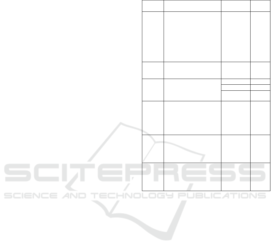
3 DATASET DESCRIPTION
The dataset, sourced from Kaggle, is organized into
two main directories: "train" and "test." Each
directory contains subdirectories, one containing X-
ray radiographs of pneumonia cases and the other
containing radiographs of normal lungs.
Specifically, 5,856 anteroposterior CXR images
from pediatric patients aged 1 to 5 years were selected
for analysis. Two labels, "pneumonia" and "normal,"
were assigned to categorize the images accordingly.
Following an adjustment and consolidation of initial
data classifications, the entire image dataset was split
into 70% for training and 30% for testing purposes.
This allocation was made to ensure a
comprehensive evaluation of the system's
performance. Consequently, 5,216 X-ray images
were allocated for training, while 640 radiographs
were reserved for testing the system's efficacy. These
images are chest X-rays (anterior-posterior) obtained
from retrospective cohorts of pediatric patients aged
one to five years old at Guangzhou Women and
Pneumonia Detection in X-Ray Chest Images Based on Convolutional Neural Networks and Data Augmentation Methods
167
Children’s Medical Center. The chest X-ray imaging
was conducted as part of the routine clinical care of
the patients.
Figure 1: Samples of the dataset.
Before inclusion in the dataset, all chest
radiographs underwent quality control screening to
eliminate any low-quality or unreadable scans.
Subsequently, the diagnoses for the images were
assessed by two expert physicians to ensure accuracy
before being used for training the AI system.
Additionally, a third expert verified the evaluation set
to address any potential grading errors, further
enhancing the reliability of the dataset.
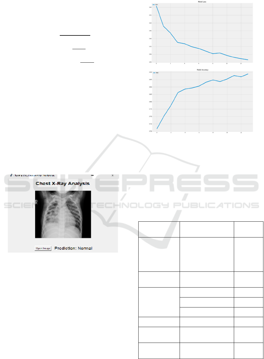
4 PROPOSED METHODOLOGY
The proposed deep learning framework has
undergone multiple constructions and training
sessions, exploring various parameters to select
optimal hyperparameters and achieve a balanced
performance architecture. Broadly, it comprises two
primary stages. The initial stage involves several
images preprocessing steps, including image resizing
to obtain a standardized size and rescaling pixel
values to fall within the [0,1] interval. Subsequently,
the second stage focuses on feature extraction and
image classification using the proposed
Convolutional Neural Network (CNN) models
(figure2).
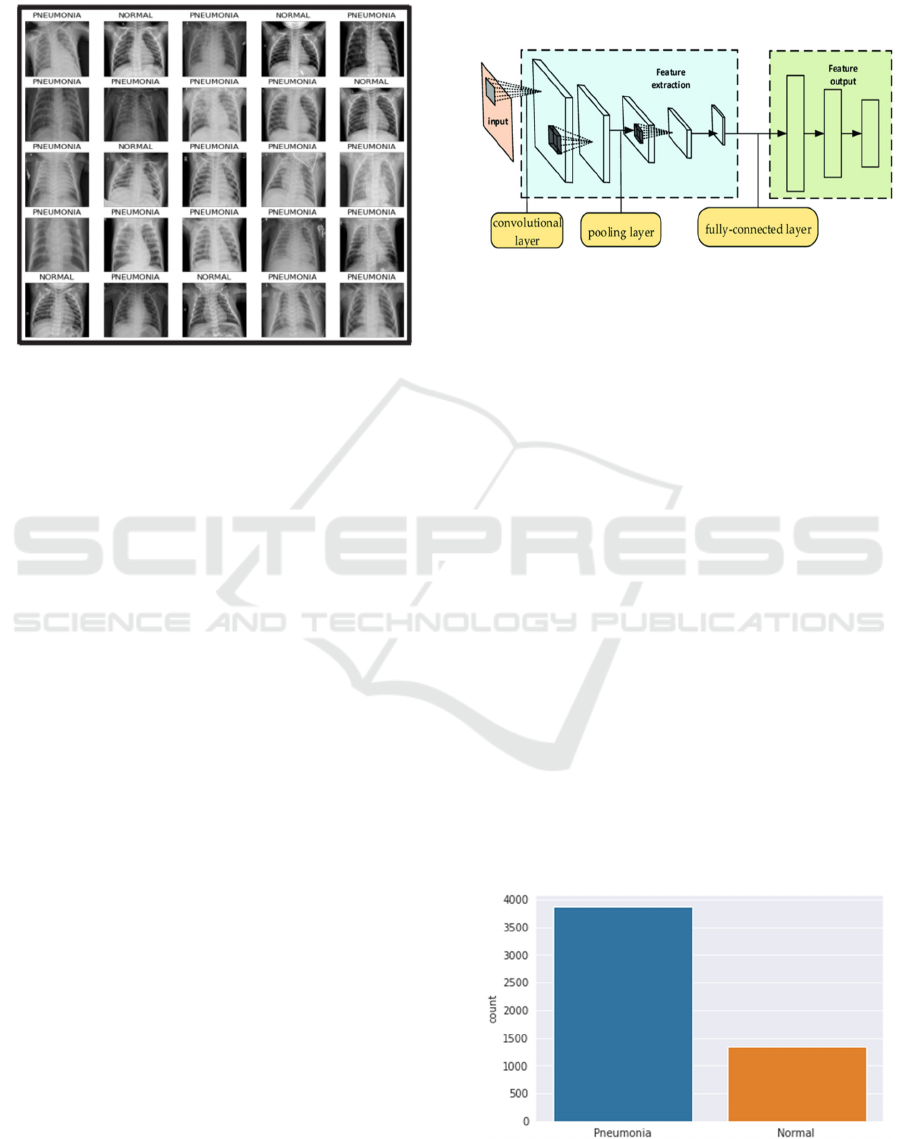
A CNN consists of multiple layers, including
convolutional layers, pooling layers, and fully
connected layers, that work together to automatically
learn spatial hierarchies of features from input images.
The convolutional layers apply filters (or kernels) to
the images to detect local patterns such as edges,
textures, and shapes. Pooling layers reduce the spatial
dimensions of the data, retaining important features
while improving computational efficiency. Finally,
the fully connected layers at the end of the network
make predictions based on the features learned by the
convolutional and pooling layers.
Figure 2: CNN structure (Sun, Shuo & Sun. 2022).
The feature extraction stage constitutes the second
component of the CNN architecture, comprising three
blocks, each containing a convolution layer,
maximum pooling layer, and dropout layer. Within
the convolutional layer, input images are transformed
into matrix representations. The convolution
operation is applied between the input matrix and a
feature kernel of a specified dimension, resulting in a
feature map. This operation effectively reduces the
dimensions of the image, facilitating further
processing. Data augmentation methods [23-24]
prove beneficial in addressing the imbalance and
scarcity of data in certain classes when dealing with
limited and uneven datasets (Figure 3). This approach
proves particularly useful for achieving a
balance in
the number of images across different MRI classes
related to brain tumors and for expanding the overall
dataset. Various augmentation techniques, including
rotation, cropping, height and width adjustments,
filling operations, zooming, and horizontal rotation
brightening, are employed to augment images and
rectify class imbalances. Given the unbalanced nature
of our dataset, this augmentation technique is applied
to artificially increase the number of images for each
class, particularly those with fewer instances.
Figure 3: Imbalanced Data.
ICT4AWE 2025 - 11th International Conference on Information and Communication Technologies for Ageing Well and e-Health
168
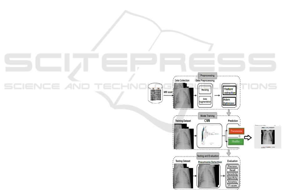
Given the apparent class imbalance in the dataset,
with potentially fewer instances of the "Pneumonia"
class compared to the "Normal" class, a strategy to
counter this issue involves leveraging data
augmentation techniques. By employing data
augmentation, we aim to augment the training dataset
by generating synthetic examples, thereby increasing
the number of instances available for training.
This approach not only addresses class imbalance
but also enhances the robustness and generalization
ability of the machine learning model. To mitigate the
risk of overfitting, expanding our dataset through
artificial means is crucial. This involves introducing
variations to the existing data via minor
transformations, thereby increasing its size.
Techniques that manipulate training data while
preserving their labels are known as data
augmentation methods . Common augmentations
include grayscale conversions, horizontal and vertical
flips, random cropping, colour adjustments,
translations, rotations, and more. By applying a
subset of these transformations to our training data,
we can substantially augment the number of
examples, leading to the creation of a highly robust
model.
For data augmentation, I implemented several
transformations to enhance the training dataset,
including randomly rotating some images by up to 30
degrees, zooming in or out by up to 20%, shifting
images horizontally by 10% of their width and
vertically by 10% of their height, and randomly
flipping images horizontally. These techniques were
applied to increase the diversity of the training data
and improve the model's ability to generalize. Once
our model is prepared, we proceed to fit the
augmented training dataset.
The imbalance in clinical
datasets, with a majority of abnormal cases and fewer
normal cases, could indeed lead to overfitting, as the
model might disproportionately favour the majority
class. To mitigate this, our study implemented data
augmentation techniques to increase the diversity and
representation of normal cases through
transformations like rotation, flipping, and scaling.
Additionally, we employed class balancing strategies,
such as adjusting class weights during training, to
penalize misclassification of the minority class more
heavily.
We validated the model using stratified cross-
validation to ensure an even class distribution across
folds and monitored evaluation metrics such as recall,
precision, and F1-score, which are sensitive to
imbalanced data. These approaches ensured the
development of a balanced and reliable model for
pneumonia detection. The proposed approach
consists of two main steps. Firstly, we introduce a
normalization method specifically designed for chest
X-rays, with the goal of removing unnecessary
components while retaining crucial information.
Following this, Deep Convolutional Neural Networks
(CNNs) are utilized, with a preference for using the
ADAM optimization function to build predictive
models using the normalized dataset.
ADAM
combines the benefits of the Adaptive Gradient
Algorithm (AdaGrad) and Root Mean Square
Propagation (RMSProp), computing adaptive
learning rates for each parameter to enhance training
efficiency and convergence Figure 4 provides an
overview of the proposed approach. The model,
consisting of 6,026,324 parameters, employs a multi-
branch convolutional architecture with three distinct
branches, each featuring different lengths and kernel
sizes to optimize feature extraction. Smaller kernels
specialize in detecting localized features such as
edges and textures, while larger kernels capture more
global patterns like shapes and contours.
By varying the branch depths, the model
combines shallow layers for basic feature recognition
with deeper layers that learn complex, high-level
abstractions. This design enables the model to process
input data at multiple scales, enriching its feature
representation and enhancing its ability to analyse
fine-grained details alongside broader patterns.
Figure 4: Proposed Architecture.
5 RESULTS AND DISCUSSION
For assessing the performance of the model, several
evaluation metrics are employed to gauge the
classification outcomes, particularly for pneumonia
Pneumonia Detection in X-Ray Chest Images Based on Convolutional Neural Networks and Data Augmentation Methods
169
classification from lung X-rays. The primary
evaluation metrics utilized include accuracy, recall
(sensitivity), and F1-score. These metrics are
calculated using the following equations:
Accuracy=
(1)
Precision=
.. (2)
Sensitivity(Recall)=
(3)
F1-score= 2/((1/𝑃𝑟𝑒𝑐𝑖𝑠𝑖on) + (1/Recall)) (4)
Here, TP denotes true positives, TN denotes true
negatives, FP denotes false positives, and FN denotes
false negatives. These metrics collectively provide a
comprehensive evaluation of the model's
performance in detection pneumonia cases from chest
X-ray images. Figure 5 illustrate view of the pneumonia
detection application. The initial step involves obtaining the
patient's information, including their name, gender, age,
phone number, history of hypertension, and the
neurologist's name. Once the user correctly fills out this
information, they are prompted to upload the lung X-ray
images. Upon submitting the chest X-Ray Images, the
application initiates the analysis using learning models to
determine the presence of a pneumonia infection.
Figure 5: Pneumonia Detection Application.
Final output of the proposed method using 12 numbers
of epoch has been shown in Figure 6. shows the model
accuracy and the corresponding loss with respect to the
number of epochs.
Our proposed model outperforms the previously
developed approaches demonstrating accuracy of
96% and the loss is 1%. Our experimental results
illustrate that the proposed CNN model exhibits
superior convergence compared to the ANN
approach, Random Forest classifier, Transfer
learning algorithms, and other CNN models. As
indicated in Table 2, our model attained the highest
accuracy rate of 96% and the best F1-Score of 94%,
along with a precision of 93%. The values attained by
our proposed CNN model stand out for their
Figure 6: The model accuracy and loss over the epochs
exceptional performance, surpassing those achieved
by the previously mentioned models. Table 2 presents
a comparison of pneumonia detection accuracy
between our proposed novel framework and state-of-
the-art models. While our proposed model achieved
accuracy that surpasses the state-of-the-art, it's
important to note that directly comparing accuracy
may not be entirely objective.
Table 2: Comparative study.
Study Method
Accuracy
Rate
Lamia A. 2022 Multilayer Perceptron
(MLP), Random
forest, Sequential
Minimal Optimization
(SMO)
84%
Shagun Sharma.
2023
Vgg16 92.15%
Jain DK. 2022 Vgg16 94%
Vgg19 95%
Xception 96%
Goyal, S. 2023 F-RNN-LSTM 95.04%
Fatma Taher.
2022
CNN 94%
Proposed model CNN+Adam
optimizer
96%
ICT4AWE 2025 - 11th International Conference on Information and Communication Technologies for Ageing Well and e-Health
170

6 CONCLUSIONS AND FUTURE
WORK
This study presents an automated method for
pneumonia detection using X ray scans, leveraging a
deep learning model for automated feature extraction
from the images. The main goal of this research was
to achieve improved classification performance with
faster learning rates compared to traditional deep
learning (DL) models. Despite the limited training
data available, experimental results demonstrate the
effectiveness of the proposed model. Its success can
be attributed to minimal preprocessing requirements
and the absence of handcrafted features, making it
suitable for diverse x ray classifications. Future
research aims to expand the classification to include
additional labels while enhancing accuracy. Future
work should aim to validate the proposed system
beyond Chest X-ray (CXR) images. It is imperative
to extend the validation to include other imaging
modalities such as computerized tomography (CT)
scans and Magnetic Resonance Imaging (MRI). This
expansion of validation will enhance the applicability
and robustness of the system across various medical
imaging techniques. Future work in pneumonia
detection using X-ray chest images could focus on the
exploration of more advanced architectures, such as
deeper or hybrid convolutional neural network
(CNN) models, which could improve detection
accuracy by capturing more complex features and
patterns. Additionally, the integration of transfer
learning from pre-trained models on large, diverse
datasets could significantly enhance performance,
particularly when labelled training data is scarce.
ACKNOWLEDGEMENTS
We would like to thank the Deanship of Scientific
Research at Shaqra University for supporting this
work.
REFERENCES
Lamia A, Fawaz A. Detection of Pneumonia Infection by
Using Deep Learning on a Mobile Platform. Comput
Intell Neurosci. 2022 Jul 30;2022:7925668. doi:
10.1155/2022/7925668. PMID: 35942467; PMCID:
PMC9356824.
Shagun Sharma, Kalpna Guleria, A Deep Learning based
model for the Detection of Pneumonia from Chest X-
Ray Images using VGG-16 and Neural Networks,
Procedia Computer Science, Volume 218, 2023.
Jain DK, Singh T, Saurabh P, Bisen D, Sahu N, Mishra J,
Rahman H. Deep Learning-Aided Automated
Pneumonia Detection and Classification Using CXR
Scans. Comput Intell Neurosci. 2022 Aug
4;2022:7474304. doi: 10.1155/2022/7474304. PMID:
35936981; PMCID: PMC9351538.
Asnake, N.W., Salau, A.O. & Ayalew, A.M. X-ray image-
based pneumonia detection and classification using
deep learning. Multimed Tools Appl (2024).
https://doi.org/10.1007/s11042-023-17965-4
Goyal, S., Singh, R. Detection and classification of lung
diseases for pneumonia and Covid-19 using machine
and deep learning techniques. J Ambient Intell Human
Comput 14, 3239–3259 (2023). https://doi.org/10.1007
/s12652-021-03464-7
Kareem, A., Liu, H. & Sant, P. Review on Pneumonia
Image Detection: A Machine Learning Approach.
Hum-Cent Intell Syst 2, 31–43 (2022). https://doi.org/
10.1007/s44230-022-00002-2
Z. Li et al., "PNet: An Efficient Network for Pneumonia
Detection," 2019 12th International Congress on Image
and Signal Processing, BioMedical Engineering and
Informatics (CISP-BMEI), Suzhou, China, 2019, pp. 1-
5, doi: 10.1109/CISP-BMEI48845.2019.8965660.
M. K. Gourisaria, V. Singh, R. Chatterjee, S. K. Panda, M.
R. Pradhan and B. Acharya, "PneuNetV1: A Deep
Neural Network for Classification of Pneumothorax
Using CXR Images," in IEEE Access, vol. 11, pp.
65028-65042, 2023, doi: 10.1109/ACCESS.2023.328
9842.
Venkateswara Reddy, E., Naveen Kumar, G.S., Siva Naga
Dhipti, G., Swathi, B. (2022). Diagnosis of Pneumonia
with Chest X-Ray Using Deep Neural Networks. In:
Reddy, V.S., Prasad, V.K., Mallikarjuna Rao, D.N.,
Satapathy, S.C. (eds) Intelligent Systems and
Sustainable Computing. Smart Innovation, Systems and
Technologies, vol 289. Springer.
Shadi A. Aljawarneh and Romesaa Al-Quraan.Pneumonia
Detection Using Enhanced Convolutional Neural
Network Model on Chest X-Ray Images. Big
Data.ahead of print http://doi.org/10.1089/big.2022.02
61.
D. S. V. Kancherla, P. Mannava, S. Tallapureddy, V.
Chintala, K. P and C. Iwendi, "Pneumothorax: Lung
Segmentation and Disease Classification Using Deep
Neural Networks," 2023 International Conference on
Self Sustainable Artificial Intelligence Systems
(ICSSAS), Erode, India, 2023, pp. 181-187, doi:
10.1109/ICSSAS57918.2023.10331853.
Farhan, A.M.Q., Yang, S. Automatic lung disease
classification from the chest X-ray images using hybrid
deep learning algorithm. Multimed Tools Appl 82,
38561–38587 (2023). https://doi.org/10.1007/s11042-
023-15047-z.
Liu, Y., Liang, P., Liang, K. et al. Automatic and efficient
pneumothorax segmentation from CT images using
EFA-Net with feature alignment function. Sci Rep 13,
15291 (2023). https://doi.org/10.1038/s41598-023-
42388-4
Pneumonia Detection in X-Ray Chest Images Based on Convolutional Neural Networks and Data Augmentation Methods
171

Chest X-Ray images (Kaggle) https://www.kaggle.com/
datasets/paultimothymooney/chest-xray-pneumonia
Khalaf Alshamrani, Hassan A. Alshamrani, Abdullah A.
Asiri, F. F. Alqahtani, Walid Theib Mohammad, Ali H.
Alshehri, "[Retracted] The Use of Chest Radiographs
and Machine Learning Model for the Rapid Detection
of Pneumonitis in Pediatric", BioMed Research
International, vol. 2022, Article ID 5260231, 11 pages,
2022.https://doi.org/10.1155/2022/5260231
Dalya S. Al-Dulaimi, Aseel Ghazi Mahmoud, Nadia
Moqbel Hassan, Ahmed Alkhayyat, Sayf A. Majeed,
"Development of Pneumonia Disease Detection Model
Based on Deep Learning Algorithm", Wireless
Communications and Mobile Computing, vol. 2022,
Article ID 2951168, 10 pages, 2022. https://doi.org/
10.1155/2022/2951168
Rajasenbagam, T., Jeyanthi, S. & Pandian, J.A. Detection
of pneumonia infection in lungs from chest X-ray
images using deep convolutional neural network and
content-based image retrieval techniques. J Ambient
Intell Human Comput (2021). https://doi.org/10.1007/
s12652-021-03075-2
Shorten, C., Khoshgoftaar, T.M. A survey on Image Data
Augmentation for Deep Learning. J Big Data 6, 60
(2019). https://doi.org/10.1186/s40537-019-0197-0
Schaudt, D., von Schwerin, R., Hafner, A. et al.
Augmentation strategies for an imbalanced learning
problem on a novel COVID-19 severity dataset. Sci
Rep 13, 18299 (2023). https://doi.org/10.1038/s41598-
023-45532-2https://doi.org/10.1155/2022/2656818
Sun, Shuo & Sun, Junzhong & Wang, Zongliang & Zhou,
Zhiyong & Cai, Wei. (2022). Prediction of Battery SOH
by CNN-BiLSTM Network Fused with Attention
Mechanism. Energies. 15. 4428. 10.3390/en15124428.
ICT4AWE 2025 - 11th International Conference on Information and Communication Technologies for Ageing Well and e-Health
172
