
Advancements in Wearable EEG Technology:
Electrode Characterization and Signal Quality Assessment
Andrea Farabbi
1 a
, Andrea Costanzo Palmisciano
1 b
, Matteo Rossi
1 c
, Niccol
`
o Antonello
2 d
,
Diana Trojaniello
2 e
, Tommaso Ongarello
2
, Pietro Cerveri
1,3 f
and Luca Mainardi
1 g
1
Department of Electronics, Information and Bioengineering, Politecnico di Milano, Milan, Italy
2
EssilorLuxottica Smart Eyewear Lab, EssilorLuxottica, 20133 Milan, Italy
3
Department of Industrial and Information Engineering, University of Pavia, 27100 Pavia, Italy
Keywords:
Electroencephalography, Wearable Systems, Biomedical Signal Processing, P300, Alpha Activity, Electrodes
Characterization.
Abstract:
This research contributes to the advancement of practical, user-friendly EEG devices for both research and
real-world applications. The paper presents a comprehensive study on the development and characterization
of wearable electroencephalography (EEG) recording in non-traditional electrode locations. In particular, we
focus on optimizing electrode placement, material selection, and signal quality assessment. Our investigation
includes impedance testing of various electrode materials, comparative analysis of dry versus wet electrodes,
and validation through standard EEG protocols. Results demonstrate the feasibility of acquiring high-quality
EEG signals from over-the-ear locations where using gold-plated brush electrodes with retractile pins, show
superior impedance characteristics (10
5
Ω) compared to other tested materials. We also validate and compare
dry electrodes by means of an eyes-open/eyes-closed protocol, confirming the ability to detect alpha rhythm
modulation in non-traditional electrode placements.
1 INTRODUCTION
Electroencephalography (EEG) has been a fundamen-
tal tool in neuroscience and clinical practice (Berger,
1929). This non-invasive technique measures the
electrical activity of the brain by recording voltage
fluctuations resulting from ionic current flows within
neurons (Teplan, 2002) and emerging to the scalp.
EEG provides excellent temporal resolution and has
a variety of application such as cognitive processes,
diagnosing neurological disorders, and developing
brain-computer interfaces (Lotte et al., 2018).
Traditional EEG systems typically utilize the in-
ternational 10-20 system for electrode placement
(Klem et al., 1999), which yields comprehensive spa-
tial information, but presents significant limitations
for its use outside controlled laboratory or clinical en-
vironments. The complexity of setup, need for skin
a
https://orcid.org/0000-0001-5582-4654
b
https://orcid.org/0009-0007-2379-1465
c
https://orcid.org/0000-0003-2519-0720
d
https://orcid.org/0000-0002-0803-5385
e
https://orcid.org/0000-0001-8935-5593
f
https://orcid.org/0000-0003-3995-8673
g
https://orcid.org/0000-0002-6276-6314
preparation, and use of conductive gels make tradi-
tional EEG systems impractical for long-term or ev-
eryday use (Casson et al., 2010).
Recent advancements in miniaturized electronics,
sensor technologies, and signal processing algorithms
have sparked interest in developing more portable and
user-friendly EEG acquisition devices (Mihajlovic
et al., 2015). These wearable EEG systems aim to
enable continuous, high-quality brain monitoring in
real-world settings, potentially expanding the applica-
tions of EEG in fields such as personalized medicine,
cognitive monitoring, and human-computer interac-
tion (Lin et al., 2014).
The development of wearable EEG technology
faces several significant challenges due to the dif-
ficulty in measuring high quality EEG signal using
small devices. These include optimizing electrode
placement for non-traditional locations, improving
signal quality from dry electrodes, ensuring user com-
fort and social acceptability, managing power con-
sumption for continuous recording, and developing
robust algorithms for real-time signal processing in
noisy environments (Mullen et al., 2015). Addition-
ally, wearable EEG systems often employ a reduced
number of channels compared to traditional setups,
which further complicates the issue of optimal elec-
716
Farabbi, A., Palmisciano, A. C., Rossi, M., Antonello, N., Trojaniello, D., Ongarello, T., Cerveri, P. and Mainardi, L.
Advancements in Wearable EEG Technology: Electrode Characterization and Signal Quality Assessment.
DOI: 10.5220/0013153300003911
Paper published under CC license (CC BY-NC-ND 4.0)
In Proceedings of the 18th International Joint Conference on Biomedical Engineering Systems and Technologies (BIOSTEC 2025) - Volume 1, pages 716-720
ISBN: 978-989-758-731-3; ISSN: 2184-4305
Proceedings Copyright © 2025 by SCITEPRESS – Science and Technology Publications, Lda.
trode placement (Debener et al., 2015). The use of dry
electrode designs is also affected by the presence of
hair which can substantially increase the impedance
potentially comprommising the measured EEG signal
quality(Lopez-Gordo et al., 2014).
In this study we chose over-the-ear locations for
EEG electrodes as it is justified by different factors.
Firstly, this area offers a balance between signal qual-
ity and user comfort, as it is less obtrusive than tra-
ditional scalp placements (Bleichner and Debener,
2017). Secondly, the proximity to temporal and pari-
etal lobes provides access to relevant brain activity
(Mikkelsen et al., 2015) while maintaining a socially
acceptable form factor (Norton et al., 2015).
This paper presents a comprehensive study ad-
dressing several key aspects of electrodes employed
in EEG enabled devices, with a specific focus on over-
the-ear electrode placement. Our research tackles
three areas of interest: i) evaluating different electrode
materials and designs to enhance signal acquisition in
this unique anatomical region; ii) comparing dry and
wet electrodes for over-the-ear placement; and iii) as-
sessing the quality of EEG signals obtained from our
over-the-ear prototype system through standard EEG
protocols and comparing them with traditional setups.
By addressing these crucial aspects in the con-
text of over-the-ear placement, our study aims to con-
tribute to the advancement of practical, user-friendly
EEG devices suitable for both research and real-world
applications.
The following sections detail our methods for
electrode characterization and signal quality assess-
ment specific to over-the-ear placement, present our
findings, and discuss their implications for the future
of wearable EEG technology.
2 METHODS
In the next sections, three data acquisitions protocols
are described and then the resulting data are later anal-
ized.
All the data acquisition on subjects was performed
according to the descriptive rules reported in the ex-
perimental protocol (Opinion 46/2023, dated Decem-
ber 18
th
, 2023) that received approval from the Po-
litecnico di Milano Ethical Committee.
2.1 Dry Impedance Testing
The study involved a set of 6 gender-balanced healthy
subjects (age: 28 ± 1.73). Four different dryelec-
trode types were tested: conductive elastomer (CE),
PLA+carbon, TPU+carbon, and gold-plated with re-
tractile pins brush (GPR) electrodes (Brainbit Inc.,
New York, NY, USA). Each electrode material was
tested three times per subject.
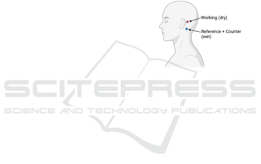
The impedance was measured using a Palm-
Sens EmStat 4S potentiostat (PalmSens BV, Houten,
Netherlands) in a two-electrode configuration. We
positioned the test electrode (serving as the working
electrode) behind the left ear, while a gel electrode,
acting as the reference and counter electrode, was
placed below the working electrode (see Figure 1 for
reference). This placement mimics the intended con-
figuration for an around-the-ear wearable EEG sys-
tem.
Figure 1: Positioning of the electrodes in the skin-electrode
impedance test.
The potentiostat generated a sine wave with an
amplitude of 100mV
rms
and 0V bias, sweeping fre-
quencies between 0.5Hz and 10kHz.
2.2 Dry vs. Wet Electrode Comparison
A set of five healthy participants (2 females, age: 28.6
± 4.84,) were recruited for a comparative study of dry
and wet electrodes during baseline recordings. For
dry electrodes, we used both brush CE and GPR elec-
trodes in occipital area. For wet electrode recordings,
we utilized an EBNeuro BEPlus LTM amplifier with
a 61-electrode cuff and gel was applied under the ex-
amined electrodes (i.e., the ones placed in the same
locations of the dry configuration).
Each subject underwent 1 minute of baseline
recording in a resting state with eyes open for each
electrode configuration.
2.3 Eyes Open/Closed Experiment
The study involved 10 healthy subjects (age: 27.2 ±
1.8, gender-balanced). We used GPR electrodes con-
nected to a MAX30001 evaluation kit (Analog De-
vices Inc., Wilmington, MA, USA), recording EEG
signals at 512Hz. Flat CE electrodes were used for
ground (GND) and reference, placed at the nasion.
Advancements in Wearable EEG Technology: Electrode Characterization and Signal Quality Assessment
717
The EOEC serves as an excellent initial test for
new EEG systems due to its simplicity and reliabil-
ity (Barry et al., 2007). The primary objectives were
to validate the ability of our chosen electrode config-
urations to capture meaningful EEG signals in both
traditional and non-traditional scalp locations, to as-
sess the sensitivity of our setup in detecting the well-
known alpha rhythm (i.e, activity in the 8 − 12Hz fre-
quency band) modulation associated with eye closure,
and to evaluate the overall signal quality and reliabil-
ity of EEG recordings obtained from our prototype
system.
Each subject participated in two recording ses-
sions:
1. GPR electrode placed in the occipital zone (serv-
ing as a ground truth for eyes-closed alpha activ-
ity)
2. GPR electrode placed over the ear (our prototype
configuration)
In each session, subjects followed a protocol alter-
nating between eyes open and eyes closed states. The
protocol consisted of two minutes with eyes open, fol-
lowed by two minutes with eyes closed. This cycle
was repeated twice, resulting in a total recording dura-
tion of eight minutes per session. The recorded signal
was then filtered between 1-30 Hz to mitigate artifacts
and line interference effects.
3 RESULTS
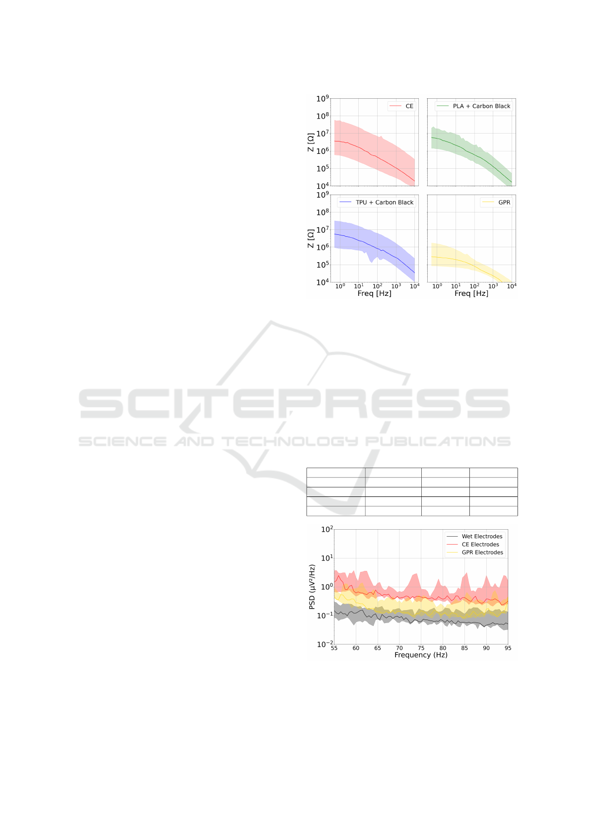
3.1 Impedance Testing
The impedance tests revealed significant differences
among the tested electrodes. GPR electrodes demon-
strated substantially lower impedance in the band
of interest (1 − 100 Hz), approximately 10
5
Ω, com-
pared to the other electrode types, which exhibited
impedance around 10
7
Ω. See Figure 2 for details on
impedance response of the materials tested.
3.2 Dry vs. Wet Electrode Comparison
In the comparison of dry and wet electrodes, we fo-
cused on the high-frequency components (above 55
Hz) of the Power Spectral Density (PSD), where noise
components are dominant. CE electrodes exhibited
the highest PSD values in this high-frequency range
(see Figure 3 for reference), indicating greater suscep-
tibility to noise. The GPR electrodes showed interme-
diate PSD values, while wet electrodes demonstrated
the lowest PSD values, confirming their superior per-
formance in noise rejection.
Figure 2: Electrode-skin impedance for the tested materials.
To further assess the signal quality, we computed
the signal prevalence for each electrode type as the
ratio of the power in each traditional brain activity
bands (delta, theta, alpha, and beta) to the power in the
high-frequency band (> 55 Hz), which we consider
representative of noise. Table 1 presents these ratios,
reported as median ± interquantile range (IQR).
Table 1: Median and IQR of brainwave band power ratios
normalized by the power in the > 55 Hz band (associated
with noise) for wet, CE, and GPR electrode types. Values
represent the relative strength of each brainwave band com-
pared to the high-frequency noise component.
Band WET GOLD DRY
delta (1-4Hz) 30.02 ± 17.07 8.49 ± 3.51 6.04 ± 5.59
theta (4-8Hz) 7.03 ± 12.33 1.52 ± 2.59 0.83 ± 1.80
alpha (8-12Hz) 2.87 ± 1.96 1.00 ± 1.29 0.36 ± 0.18
beta (12-30Hz) 2.18 ± 1.86 0.85 ± 0.99 0.61 ± 0.27
Figure 3: Comparison of Power Spectral Density (PSD) for
frequencies above 55Hz between CE, GPR, and wet elec-
trodes. PSD is reported on a logarithmic scale, with median
IQR shown for each electrode type.
BIOSIGNALS 2025 - 18th International Conference on Bio-inspired Systems and Signal Processing
718
In this analysis, higher values correspond to more
reliable and robust signals. Consistent with the PSD
results, wet electrodes achieved the highest ratios
across all frequency bands, indicating the best sig-
nal quality. The GPR electrodes showed intermedi-
ate performance, while the CE electrodes yielded the
lowest ratios, suggesting they are more susceptible to
noise interference. Due to the superior impedance
characteristics demonstrated by GPR electrodes, the
subsequent EOEC experimental results will focus ex-
clusively on this electrode type
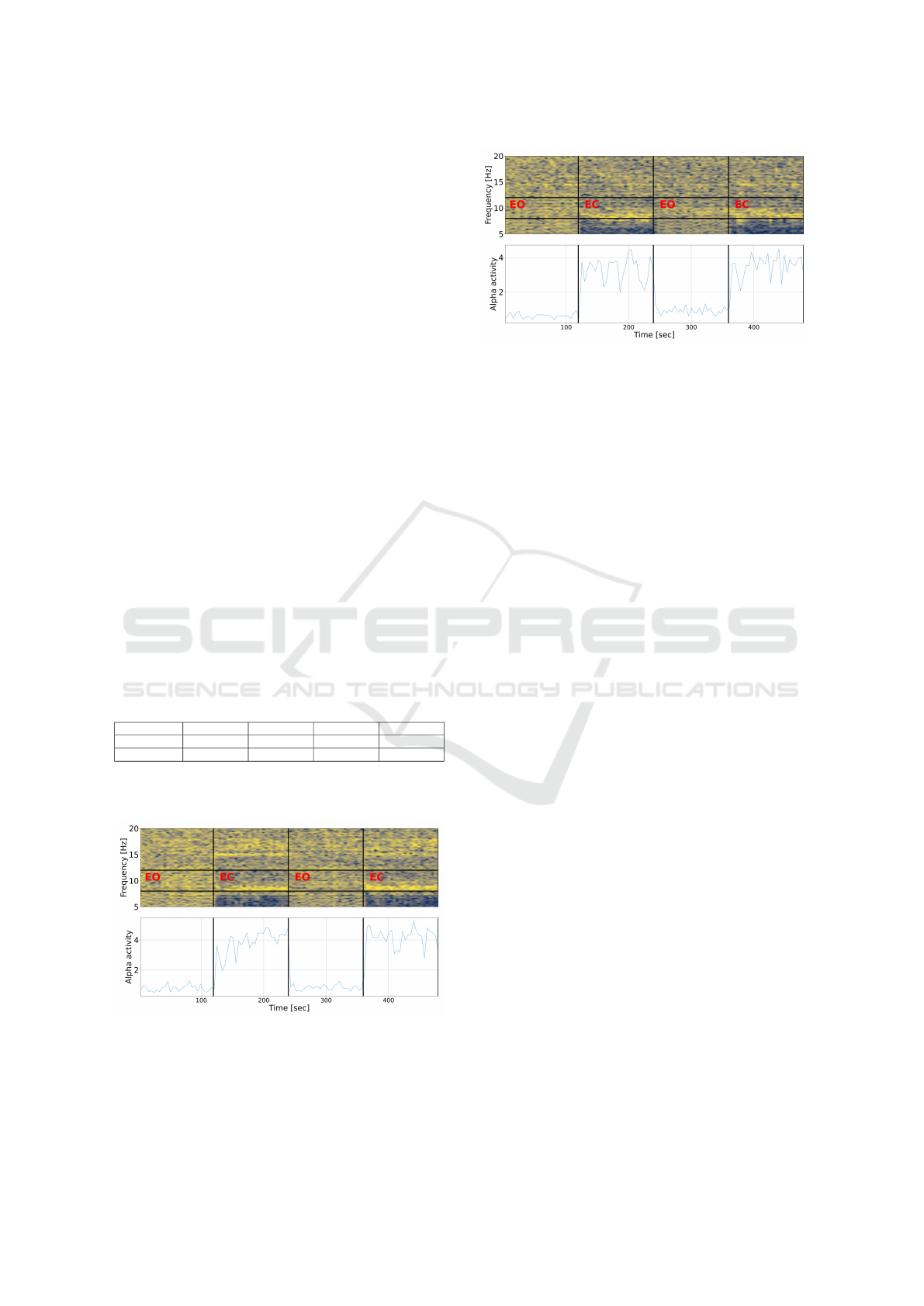
3.3 Eyes Open/Closed Experiment
Both electrode placements (occipital and over-ear)
captured clear modulation of alpha activity corre-
sponding to the eyes open/closed cycles. The spectro-
grams (see Figures 4 and 5) showed increased power
in the alpha frequency band (8-12 Hz) during eyes-
closed periods as expected (Barry et al., 2007). This
effect was more pronounced in the occipital electrode
placement (as expected from the literature), but was
also clearly visible in the over-the-ear configuration.
The alpha ratio (power in alpha normalized
by broadband power) analysis revealed significantly
higher values during the eyes-closed phases in both
recording setups (see Table 2).
Table 2: Distribution of Alpha ratio recorded in the occipital
and over-the-ear locations in the different phases of the pro-
tocols (EO: eyes open, EC: eyes closed) along the examined
population (median ± IQR).
Location EO 1 EC 1 EO 2 EC 2
Occipital 0.76 ± 0.21 3.88 ± 0.74 0.81 ± 0.17 4.21 ± 0.60
Over-the-ear 0.62 ± 0.13 3.31 ± 0.67 0.92 ± 0.20 3.54 ± 0.59
The results show that both electrode placements
successfully captured the expected alpha rhythm
Figure 4: Spectrogram (top) and alpha ratio (bottom) for the
occipital electrode placement during eyes open/closed cy-
cles. The spectrogram show power across frequencies over
time, while the alpha ratio plot displays the ratio of alpha
band power to total power. EO = Eye-Open period; EC =
Eye-Closed period.
Figure 5: Spectrogram (top) and alpha ratio (bottom) for
the over-ear electrode placement during eyes open/closed
cycles. The spectrogram shows power across frequencies
over time, while the alpha ratio plot displays the ratio of
alpha band power to total power. EO = Eye-Open period;
EC = Eye-Closed period.
modulation between eyes-open and eyes-closed con-
ditions. As anticipated, the occipital placement
showed the strongest effect, with the highest alpha
ratios during eyes-closed periods. The over-the-ear
placement demonstrated a robust ability to detect the
alpha rhythm modulation, with alpha ratios compara-
ble to those observed in the occipital placement.
These findings suggest that the over-the-ear elec-
trode placement, using GPR electrodes, is capable of
reliably detecting fundamental EEG phenomena such
as alpha rhythm modulation. This supports the po-
tential of this configuration for use in wearable EEG
devices.
4 DISCUSSION
Our study on wearable over-the-ear EEG technol-
ogy revealed several significant findings. GPR elec-
trodes demonstrated superior impedance characteris-
tics (10
5
Ω compared to 10
7
Ω for other types), sug-
gesting better electrode-skin interface performance.
While wet electrodes showed the best signal quality,
GPR electrodes provided a practical alternative bal-
ancing signal quality and user convenience.
The EOEC experiments validated the feasibility
of acquiring meaningful EEG signals from over-the-
ear locations, with alpha activity modulation compa-
rable to traditional occipital placements. This sug-
gests over-the-ear placement could be viable for cer-
tain EEG applications, offering advantages in user
comfort and social acceptability. The ability to cap-
ture meaningful EEG data from this location is par-
ticularly significant for developing wearable devices
that could be worn for extended periods in everyday
settings.
Advancements in Wearable EEG Technology: Electrode Characterization and Signal Quality Assessment
719

The combination of over-the-ear electrode place-
ment and GPR electrodes offers a promising config-
uration for wearable EEG devices, providing an opti-
mal balance of signal quality, potential user comfort,
and practical applicability. This addresses key chal-
lenges in developing wearable EEG technology for
everyday use. However, while our results are promis-
ing for short-term recordings, the long-term stability
and comfort of the proposed configurations require
further investigation. Additionally, performance dur-
ing physical activity or in noisy environments needs
to be assessed.
Future work should focus on increasing sample
size, assessing long-term stability and comfort, inves-
tigating performance during complex cognitive tasks
beyond the EOEC paradigm, and developing spe-
cialized signal processing algorithms for over-the-ear
recordings. These investigations will provide a more
comprehensive understanding of the capabilities and
limitations of over-the-ear EEG recordings.
5 CONCLUSION
Our research demonstrated that GPR electrodes
achieve superior impedance characteristics for over-
the-ear EEG signal acquisition. The successful de-
tection of alpha rhythm modulation in over-the-ear
locations, comparable to traditional occipital place-
ment, validates this approach for EEG recording. The
combination of over-the-ear placement and GPR elec-
trodes provides a promising configuration for wear-
able EEG devices, effectively balancing signal quality
and user comfort. Future studies investigating more
complex cognitive tasks beyond EOEC paradigms
would further validate and strengthen these findings,
potentially expanding the applications of this wear-
able EEG technology.
ACKNOWLEDGEMENTS
This work was carried out in the EssilorLuxottica
Smart Eyewear Lab, a Joint Research Center between
EssilorLuxottica and Politecnico di Milano.
REFERENCES
Barry, R. J., Clarke, A. R., Johnstone, S. J., Magee, C. A.,
and Rushby, J. A. (2007). Eeg differences between
eyes-closed and eyes-open resting conditions. Clinical
Neurophysiology, 118(12):2765–2773.
Berger, H. (1929).
¨
Uber das elektrenkephalogramm
des menschen. Archiv f
¨
ur psychiatrie und ner-
venkrankheiten, 87(1):527–570.
Bleichner, M. G. and Debener, S. (2017). Concealed, un-
obtrusive ear-centered eeg acquisition: ceegrids for
transparent eeg. Frontiers in Human Neuroscience,
11.
Casson, A. J., Yates, D. C., Smith, S. J., Duncan, J. S., and
Rodriguez-Villegas, E. (2010). Wearable electroen-
cephalography. IEEE engineering in medicine and bi-
ology magazine, 29(3):44–56.
Debener, S., Emkes, R., de Vos, M., and Bleichner, M.
(2015). Unobtrusive ambulatory eeg using a smart-
phone and flexible printed electrodes around the ear.
Scientific Reports, 5.
Klem, G. H., L
¨
uders, H., Jasper, H. H., and Elger, C. E.
(1999). The ten-twenty electrode system of the in-
ternational federation. the international federation of
clinical neurophysiology. Electroencephalography
and clinical neurophysiology. Supplement, 52:3–6.
Lin, C.-T., Chuang, C.-H., Wang, Y.-K., Tsai, S.-F., Chiu,
T.-P., and Ko, L.-W. (2014). Wireless and wear-
able eeg system for evaluating driver vigilance. IEEE
Transactions on Biomedical Circuits and Systems,
8(2):165–176.
Lopez-Gordo, M. A., Sanchez-Morillo, D., and Valle, F. P.
(2014). Dry eeg electrodes. Sensors, 14(7):12847–
12870.
Lotte, F., Bougrain, L., Cichocki, A., Clerc, M., Con-
gedo, M., Rakotomamonjy, A., and Yger, F. (2018).
A review of classification algorithms for EEG-based
brain–computer interfaces: a 10 year update. Journal
of neural engineering, 15(3):031005.
Mihajlovic, V., Grundlehner, B., Vullers, R., and Penders, J.
(2015). Wearable, wireless eeg solutions in daily life
applications: what are we missing? IEEE journal of
biomedical and health informatics, 19(1):6–21.
Mikkelsen, K. B., Kappel, S. L., Mandic, D. P., and Kid-
mose, P. (2015). Eeg recorded from the ear: Char-
acterizing the ear-eeg method. Frontiers in Neuro-
science, 9.
Mullen, T. R., Kothe, C. A., Chi, Y. M., Ojeda, A., Kerth, T.,
Makeig, S., Jung, T.-P., and Cauwenberghs, G. (2015).
Real-time neuroimaging and cognitive monitoring us-
ing wearable dry eeg. IEEE Transactions on Biomed-
ical Engineering, 62(11):2553–2567.
Norton, J., Lee, D. S., Lee, J., Lee, W., Kwon, O., Won, P.,
Jung, S., Cheng, H., Jeong, J.-W., Akce, A., Umunna,
S., Na, I., Kwon, E. Y. H., Wang, X.-Q., Liu, Z., Paik,
U., Huang, Y., Bretl, T., and Rogers, J. (2015). Soft,
curved electrode systems capable of integration on the
auricle as a persistent brain–computer interface. Pro-
ceedings of the National Academy of Sciences of the
United States of America, 112.
Teplan, M. (2002). Fundamentals of EEG measurement.
Measurement science review, 2(2):1–11.
BIOSIGNALS 2025 - 18th International Conference on Bio-inspired Systems and Signal Processing
720
