
Webcam-Based Pupil Diameter Prediction Benefits from Upscaling
Vijul Shah
1 a
, Brian B. Moser
1,2 b
, Ko Watanabe
1,2 c
and Andreas Dengel
1,2 d
1
RPTU Kaiserslautern-Landau, Germany
2
German Research Center for Artificial Intelligence (DFKI), Germany
fi
Keywords:
Pupil Diameter Prediction, Image Super-Resolution.
Abstract:
Capturing pupil diameter is essential for assessing psychological and physiological states such as stress levels
and cognitive load. However, the low resolution of images in eye datasets often hampers precise measure-
ment. This study evaluates the impact of various upscaling methods, ranging from bicubic interpolation to
advanced super-resolution, on pupil diameter predictions. We compare several pre-trained methods, including
CodeFormer, GFPGAN, Real-ESRGAN, HAT, and SRResNet. Our findings suggest that pupil diameter pre-
diction models trained on upscaled datasets are highly sensitive to the selected upscaling method and scale.
Our results demonstrate that upscaling methods consistently enhance the accuracy of pupil diameter predic-
tion models, highlighting the importance of upscaling in pupilometry. Overall, our work provides valuable
insights for selecting upscaling techniques, paving the way for more accurate assessments in psychological
and physiological research.
1 INTRODUCTION
The widespread adoption of eye-tracking technology
in daily life is accelerating, as highlighted by innova-
tions like Apple’s camera-based eye tracking (Apple
Inc., 2024), (Greinacher and Voigt-Antons, 2020).
As a fortunate side-effect, these technologies en-
able the analysis of human cognitive states, which
are deeply connected to observable features in the
eyes (Dembinsky et al., 2024a), (Dembinsky et al.,
2024b). While much of the existing research focuses
on blink detection (Hong et al., 2024) and gaze es-
timation (O’Shea and Komeili, 2023), (Yun et al.,
2022), (Bhatt et al., 2024), which employ biomarker
usage (Liu et al., 2022), infrared reflections (Fathi
and Abdali-Mohammadi, 2015), or image analysis
techniques (Hisadome et al., 2024), there is com-
paratively less emphasis on measuring pupil diame-
ters (Sari et al., 2016), (Caya et al., 2022). Yet,
accurately capturing pupil size is critical for assess-
ing various physiological and psychological condi-
tions: Recent research shows that the diameter of
the pupil can indicate levels of stress (Pedrotti et al.,
2014), focus (L
¨
udtke et al., 1998), (Van Den Brink
a
https://orcid.org/0009-0008-5174-0793
b
https://orcid.org/0000-0002-0290-7904
c
https://orcid.org/0000-0003-0252-1785
d
https://orcid.org/0000-0002-6100-8255
et al., 2016), or cognitive load (Kahneman and Beatty,
1966), (Pfleging et al., 2016), (Krejtz et al., 2018).
Moreover, pupil size is linked to the activity of the
locus coeruleus (Murphy et al., 2014), (Joshi et al.,
2016), a crucial brain region for memory manage-
ment over both short and long terms (Kahneman and
Beatty, 1966), (Kucewicz et al., 2018). It is also
vital in other medical contexts, such as evaluating
the pupillary responses relating to neurological con-
ditions like Alzheimer’s disease (Granholm et al.,
2017), (Tales et al., 2001), (Kremen et al., 2019),
schizophrenia (Reddy et al., 2018), Parkinson’s dis-
ease (Micieli et al., 1991), opioid use (Murillo
et al., 2004), mild cognitive impairment (Elman et al.,
2017), and in patients with brain injuries in intensive
care settings (Kotani et al., 2021). Therefore, precise
estimation of pupil diameter is essential for advancing
the effectiveness of image-based eye-tracking tech-
nologies.
The introduction of the EyeDentify (Shah et al.,
2024) dataset, which offers webcam-based eye im-
ages with corresponding pupil diameters, marks a sig-
nificant advancement in pupilometry research. Un-
like previous datasets (Ni and Sun, 2019), (Khokhlov
et al., 2020) that were either not publicly accessible
or recorded under highly controlled conditions, Eye-
Dentify provides a diverse array of recordings fea-
turing varying seating positions and distances. Thus
376
Shah, V., Moser, B. B., Watanabe, K. and Dengel, A.
Webcam-Based Pupil Diameter Prediction Benefits from Upscaling.
DOI: 10.5220/0013162800003890
In Proceedings of the 17th International Conference on Agents and Artificial Intelligence (ICAART 2025) - Volume 2, pages 376-385
ISBN: 978-989-758-737-5; ISSN: 2184-433X
Copyright © 2025 by Paper published under CC license (CC BY-NC-ND 4.0)

potentially advancing the development of consumer-
grade pupillometers that are capable of handling di-
verse eye colors and are easily accessible without any
significant efforts, position constraints, and technical
expertise. However, the primary challenge with this
dataset is the low quality of the images, which can
be attributed to the recording camera quality and the
small size of the eyes within the images. This neces-
sitates the application of image upscaling techniques
to enable the effective use of deep neural networks for
pupil diameter prediction.
In this work, we explore the impact of various
image Super-Resolution (SR) techniques on the ac-
curacy of webcam-based pupil diameter predictions.
Image SR aims to transform low-resolution images
into high-resolution counterparts, potentially enhanc-
ing the clarity and detail of visual data used in train-
ing models for more accurate pupil diameter estima-
tion (Moser et al., 2023). We demonstrate that em-
ploying advanced, pre-trained SR models can sub-
stantially improve the accuracy of pupil diameter pre-
dictions in low-quality, webcam-based images. Yet,
we found that different image SR methods affect pupil
diameter estimation differently. The effectiveness of
SR methods varied, with some enhancing the features
necessary for precise pupilometry more effectively
than others. Nevertheless, we can conclude that using
upscaling methods, in general, improves the perfor-
mance of pupil diameter prediction models. Overall,
our comparative analysis provides clear guidance on
selecting appropriate SR techniques for pupilometry.
2 RELATED WORK
In this section, we briefly review the usage of image
SR as a pre-processing step for downstream tasks and
survey the state-of-the-art of pupil diameter estima-
tion.
2.1 Super-Resolution as Pre-Processing
Image SR is the process of transforming a LR im-
age into a HR one, effectively solving an inverse
problem (Moser et al., 2023). More explicitly, a SR
model M
θ
: R
H×W×C
→ R
s·H×s·W×C
is trained to in-
verse the degradation relationship between a LR im-
age x ∈ R
H×W×C
and the HR image y ∈ R
s·H×s·W×C
,
where s denotes the scaling factor and the degradation
relationship can be described by
x = ((y ⊗ k) ↓
s
+n)
JPEG
q
, (1)
where k is a blur kernel, n the additive noise, and q
the quality factor of a JPEG compression. In a su-
pervised setting, the training is based on a dataset
D
SR
= {(x
i
, y
i
)}
N
i=1
of LR-HR image pairs of cardi-
nality N and on the overall optimization target
θ
∗
= argmin
θ
E
(x
i
,y
i
)∈D
SR
∥M
θ
(x
i
) − y
i
∥
2
(2)
Trained SR models are utilized across a wide array
of fields, enhancing everything from medical imag-
ing, where increased image clarity can have criti-
cal implications for patient care, to satellite imagery
that provides more detailed insights into Earth’s ge-
ography (Song et al., 2022), (Tang et al., 2021).
In consumer electronics, such as smartphones and
high-definition televisions, SR technologies signifi-
cantly improve the visual quality, creating more en-
gaging and realistic digital experiences (Zhan et al.,
2021), (Shi et al., 2016). With the rapid advance-
ments driven by deep learning and cutting-edge gen-
erative models, the field of image SR has experienced
significant progress (Moser et al., 2024b), (Li et al.,
2023), (Bashir et al., 2021). This work, however, does
not seek to develop new image SR methodologies. In-
stead, it leverages SR technology as a preprocessing
step to enhance the precision of pupil diameter mea-
surements for images in everyday settings.
Similar applications of pre-trained SR models for
downstream tasks inspire our goal in related fields,
such as image recognition (Kim et al., 2024), (He
et al., 2024), remote sensing (Chen et al., 2024),
dataset distillation (Moser et al., 2024a), and oth-
ers (Liu, 2024), (Jiang et al., 2024). For instance,
Chen et al. utilized image SR to improve the qual-
ity of semantic segmentation (Chen et al., 2023a). In
a different context, Mustafa et al. adopted image SR
as a defensive strategy against adversarial attacks on
image classification systems (Mustafa et al., 2019).
Similarly, Na et al. applied image SR to boost the
performance of object classification algorithms (Na
and Fox, 2020). By integrating image SR into our
workflow, we aim to refine the input data quality, thus
enabling more accurate and reliable analyses in pupil
diameter estimation.
2.2 Pupil Diameter Estimation
Ni et al. introduced a method named BINOMAP
for estimating pupil diameter, utilizing dual cameras
- referred to as master and slave - as a binocular
geometric constraint for analyzing gaze images (Ni
and Sun, 2019). This model is built on Zhang’s al-
gorithm, which recorded a mean absolute error of
0.022 ± 0.017mm (Zhang, 1999). Similarly, Caya et
al. used a camera positioned 10cm away from the
subject’s face to capture facial images. These im-
ages were then processed on a Raspberry Pi, which
involved converting RGB images to grayscale, adjust-
Webcam-Based Pupil Diameter Prediction Benefits from Upscaling
377
Cropped Eyes
(64 x 32)
Eye Extraction
SR Image
(1280 x 960)
Original
(640 x 480)
Super-
Resolution
Face Crop
(512 x 512)
Face
Landmarks
Save as pupil
diameter data
Blink Removal
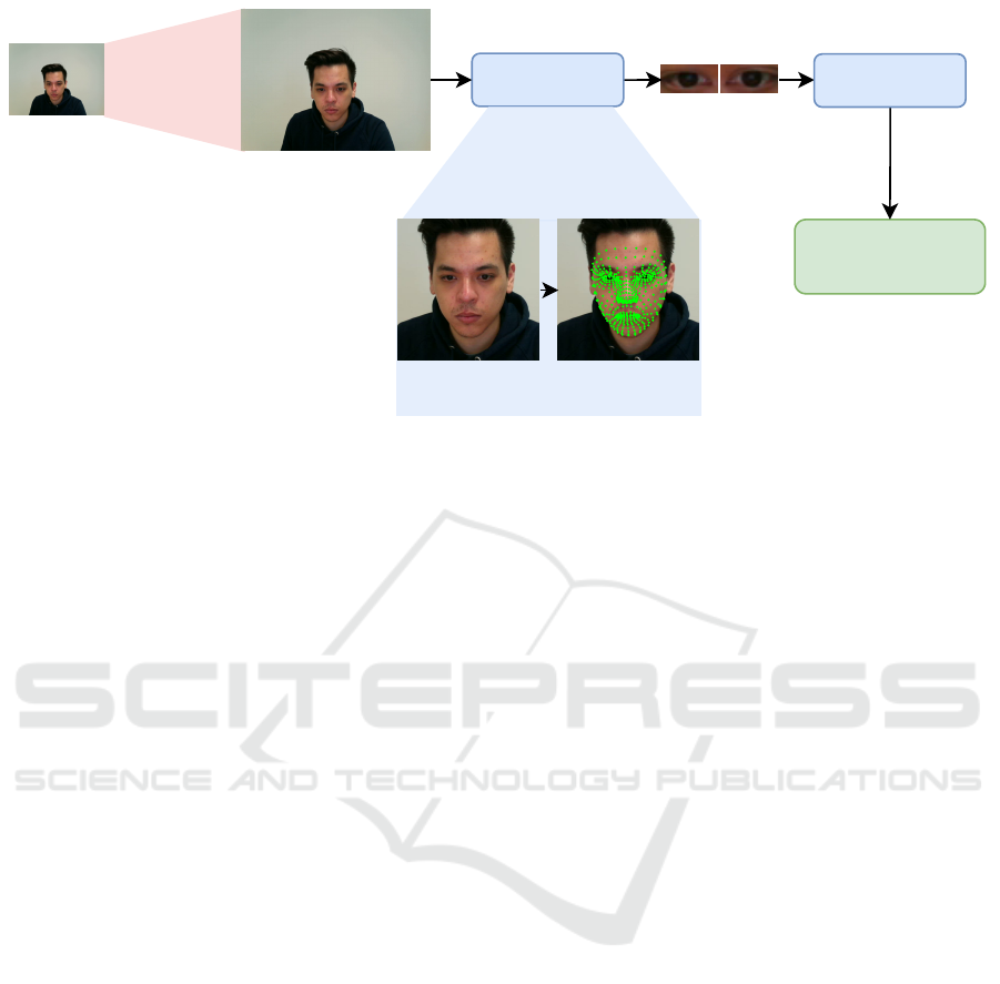
Figure 1: Pipeline of our data preprocessing with image SR. As a first step, we super-resolve the raw data with a pre-defined
scaling factor (here 2×). Next, we used Mediapipe to extract the respective cropped eye images (64 × 32), left and right, for
face detection and landmark localization. Subsequently, we applied blink detection on the cropped eyes using the Eye Aspect
Ratio (EAR) and a pre-trained vision transformer for blink detection, as described in EyeDentify (Shah et al., 2024). Cropped
eye images are then saved based on the EAR threshold and model confidence score.
ing contrast and brightness, reshaping images, and
applying the Tiny-YOLO algorithm for pupil diam-
eter estimation (Khokhlov et al., 2020). Their ap-
proach resulted in measurement accuracies with a per-
cent difference of 0.58% for the left eye and 0.48% for
the right eye. Both works face significant constraints
related to specific conditions, including the necessity
for dual cameras and maintaining a constant, fixed
distance between the face and the camera. Another
major limitation of these works is that their datasets
are not publicly available, contrary to the EyeDentify
dataset (Shah et al., 2024).
3 METHODOLOGY
The goal of this work is to apply SR models of the
form M
θ
: R
H×W×C
→ R
s·H×s·W×C
to improve the
quality of eye images derived from face webcam im-
ages, denoted as D
eyes
⊂ D
f aces
, which is crucial
for accurate pupil diameter estimation and cognitive
state analysis. More formally, we aim at constructing
D
M
θ
eyes
= {(M
θ
(
ˆ
x
i
), y
i
)}
N
i=1
, where (
ˆ
x
i
, y
i
) ∈ D
eyes
⊂
R
H×W×C
× R,
ˆ
x
i
∈ R
H×W×C
denotes the webcam im-
ages of eyes and y
i
∈ R their respective pupil diame-
ter size. Due to the sparsity of available training data
in this eye-monitoring domain (Shah et al., 2024),
we primarily refer to pre-trained SR models with
given parameters θ instead of training a model M
θ
from scratch. Figure 1 illustrates the overall pipeline,
which integrates SR, i.e., M
θ
, before any face detec-
tion, eye localization, cropping, and blink detection.
This revised methodology leverages the strengths of
existing SR models while tailoring their application
to meet the specific demands of eye feature analysis.
Initially, we planned to apply pre-trained SR tech-
niques directly to isolated images of the left and right
eyes, as suggested by the authors of EyeDentify (Shah
et al., 2024). However, this approach faces significant
limitations, such as the rarity of eye images in im-
age SR training datasets, e.g., DIV2K (Agustsson and
Timofte, 2017) or Flicker2K (Timofte et al., 2017).
State-of-the-art SR models like HAT (Chen et al.,
2023b) or face SR models like GFPGAN (Wang et al.,
2021a) are primarily optimized for everyday or full-
face images. When these models are applied directly
to eye images, their effectiveness diminishes due to
a mismatch in the data distribution and latent space,
which are tailored for the complexities of everyday or
entire face features, as shown in Figure 2. To address
this issue, we propose a more general approach: in-
stead of applying SR directly to eye webcam images
D
M
θ
eyes
⊂ D
M
θ
f aces
, we utilize the entire face webcam im-
ages D
M
θ
f aces
. Thus, our revised goal is to derive
D
M
θ
f aces
= {(M
θ
(x
i
), y
i
)}
N
i=1
, (3)
where x
i
∈ D
f aces
⊂ R
H×W×C
denotes the web-
cam full-face images before any eye-cropping g :
R
H×W×C
→ R
H
′
×W
′
×C
with H
′
≪ H and W
′
≪ W
happened, i.e.,
ˆ
x
i
= g (x
i
). This allows the SR models
trained on classical SR datasets D
SR
to operate within
ICAART 2025 - 17th International Conference on Agents and Artificial Intelligence
378

Figure 2: Comparison of applying image SR models on the cropped eye images versus applying them on the entire image.
While the SR approximations on the entire image lead to results plausible to the respective input, the SR models applied to
the cropped eye images lead to very distinct images. For instance, GFPGAN (left) produces unnatural pupils, whereas HAT
(right) emits brightness shifts.
their optimal data distribution context, i.e.,
∥µ
D
SR
− µ
D
f aces
∥
2
≪ ∥µ
D
SR
− µ
D
eyes
∥
2
and
Tr
Σ
D
SR
+ Σ
D
f aces
− 2
q
Σ
D
SR
Σ
D
f aces
≫ Tr
Σ
D
SR
+ Σ
D
eyes
− 2
q
Σ
D
SR
Σ
D
eyes
,
where Tr(·) denotes the trace of a matrix, µ
(·)
the
means and Σ
(·)
the respective covariances. After en-
hancing the overall facial images, we proceed with
localized feature extraction focused on the eyes. This
includes precise eye localization, cropping, and sub-
sequent analyses such as blink detection, which we
can describe as a function ϕ
blink
such that |D
M
θ
eyes
| ≫
|ϕ
blink
D
M
θ
eyes
|.
3.1 SR Techniques
Regarding SR methodologies, we identify two pri-
mary factors that fundamentally influence the perfor-
mance and outcomes of SR models M
θ
: the architec-
ture of the models and their training objectives to opti-
mize θ (Moser et al., 2024b). Based on the latter, SR
models can be broadly categorized into two groups:
regression-based models, which typically employ a
regression loss, and generative SR models, which uti-
lize adversarial loss mechanisms. These distinctions
are crucial as they result in varying SR approxima-
tions, which can subsequently impact the accuracy of
pupil diameter estimations. To encompass the breadth
of techniques available and ensure a comprehensive
evaluation, we have selected at least two distinct ap-
proaches from each category:
• Regression-Based Models.
– SRResNet. A general SR method that draws
architectural inspiration from ResNet (He et al.,
2016; Ledig et al., 2017).
– HAT. A state-of-the-art vision transformer de-
signed for image SR (Dosovitskiy et al., 2020;
Chen et al., 2023b).
• Generative Models.
– GFPGAN. A face-oriented SR GAN model de-
signed specifically to enhance facial features
within images (Wang et al., 2021b).
– CodeFormer. A face-oriented VQ-VAE based
model (Zhou et al., 2022).
– Real-ESRGAN. A more generalized SR GAN
approach, which is considered to offer robust
solutions for generating photorealistic textures
and details in everyday situations (Wang et al.,
2022).
3.2 EyeDentify++
As a result of the examination of GFPGAN, Code-
Former, Real-ESRGAN, HAT, and SRResNet SR
models for pupil diameter estimation, we can cre-
ate five additional datasets containing left and right
eye images separately, which we call EyeDen-
tify++
1
. Due to the different SR approximations,
the later stages, where we recognize faces, crop eyes,
and detect blinks, result in retaining and discard-
ing different amounts of images. More formally,
|ϕ
blink
D
GFPGAN×2
eyes
| ̸= |ϕ
blink
D
HAT ×2
eyes
|. Figure 3
compares the number of images in the original dataset
with those in the SR datasets after blink detection.
The results indicate that SR enhances the accuracy
of blink classification by improving the calculation
of the EAR ratio through clearer eye landmark detec-
tion on the 2x and 4x up-scaled images and provid-
ing higher-quality images for feature extraction in the
subsequent blink detection phase (Shah et al., 2024).
1
https://vijulshah.github.io/webcam-based-pupil-
diameter-estimation/
Webcam-Based Pupil Diameter Prediction Benefits from Upscaling
379

Figure 3: Comparison of applying pre-trained SR models on the EyeDentify Dataset.
4 EXPERIMENTS
In this section, we present our experimental part,
which consists of model and training details as well
as quantitative and qualitative results.
4.1 Model Details
For pupil diameter prediction, we employed the same
regression models as suggested in EyeDentify (Shah
et al., 2024): ResNet18, ResNet50, and ResNet152,
with the same model configuration and processing
steps. The datasets created through SR methods were
used to train and evaluate these ResNet models. We
upscaled the eye images by 2x and 4x using bi-cubic
interpolation to reach 64 x 32 and 128 x 64 dimen-
sions. We then refined the images using SR models
(e.g., GFPGAN, CodeFormer, Real-ESRGAN, HAT,
and SRResNet).
4.2 Training Details
We followed the training setup from the original work
(Shah et al., 2024). Using 5-fold cross-validation,
we trained ResNet18, ResNet50, and ResNet152 from
scratch on all datasets for 50 epochs, with a batch size
of 128, separately for left and right eyes. We used
the AdamW optimizer with default settings, a weight
decay of 10
−2
, and an initial learning rate of 10
−4
,
which was reduced by 0.2 every 10 epochs.
5 RESULTS
Table 1 presents 5-fold cross-validation results for
ResNet18, ResNet50, and ResNet152 on SRx2 and
SRx4 datasets. Compared to the original EyeDentify
dataset, we can observe that upscaling greatly benefits
pupil diameter prediction.
Scale Sensitivity. Table reveals a complex relation-
ship between the scale factor and the performance of
SR methods. There is no consistent trend of improve-
ment or deterioration as the scale increases from ×2
to ×4 across all methods.
Potential Overfitting. Certain SR methods exhibit
exceptional performance in specific configurations
but perform poorly in others. For instance, while
ResNet152 shows improved results with bicubic in-
terpolation at ×2 scale, it tends to overfit with SR at
higher scales. This variability could indicate overfit-
ting to particular network architectures, highlighting a
need for robustness in classifier selection rather than
focusing solely on image enhancement.
Best Models. Across different setups, bicubic up-
sampling frequently achieves optimal performance
for both left and right eyes, particularly notable in
the ResNet18 architecture. However, advanced SR
methods like Real-ESRGAN and SRResNet also con-
sistently demonstrate lower error rates, underscoring
their potential effectiveness in specific configurations.
These findings suggest a balanced approach in select-
ing SR methods, considering both traditional tech-
niques and advanced models based on specific needs.
Visualizations. Figure 5 shows the Class Activation
Maps (CAM) (Zhou et al., 2016) from the final con-
volution layer for each model, tested on a participant
viewing the same display color across all datasets.
ICAART 2025 - 17th International Conference on Agents and Artificial Intelligence
380

Table 1: Quantitative Mean Absolute Error (MAE) ↓ comparison across different pre-trained SR methods and pupil diameter
prediction models for both left and right eyes. The lowest errors are highlighted.
Eye Scale Method ResNet18 ResNet50 ResNet152
Left
×1 No SR 0.1329 ± 0.0235 0.1280 ± 0.0164 0.1259 ± 0.0176
×2
Bi-cubic 0.1340 ± 0.0196 0.1402 ± 0.0327 0.1225 ± 0.0166
GFPGAN 0.1428 ± 0.0360 0.1486 ± 0.0195 0.1339 ± 0.0122
CodeFormer 0.1328 ± 0.0245 0.1476 ± 0.0364 0.1442 ± 0.0189
Real-ESRGAN 0.1265 ± 0.0179 0.1369 ± 0.0153 0.1384 ± 0.0195
SRResNet 0.1286 ± 0.0139 0.1249 ± 0.0062 0.1391 ± 0.0261
HAT 0.1251 ± 0.0129 0.1277 ± 0.0241 0.1418 ± 0.0197
×4
Bi-cubic 0.1375 ± 0.0192 0.1382 ± 0.0287 0.1497 ± 0.0275
GFPGAN 0.1397 ± 0.0244 0.1230 ± 0.0122 0.1348 ± 0.0183
CodeFormer 0.1383 ± 0.0170 0.1404 ± 0.0201 0.1413 ± 0.0164
Real-ESRGAN 0.1338 ± 0.0178 0.1306 ± 0.0160 0.1316 ± 0.0183
SRResNet 0.1384 ± 0.0234 0.1345 ± 0.0163 0.1509 ± 0.0242
HAT 0.1330 ± 0.01191 0.1305 ± 0.0115 0.1454 ± 0.0179
Right
×1 No SR 0.1548 ± 0.0273 0.1501 ± 0.0214 0.1452 ± 0.0163
×2
Bi-cubic 0.1402 ± 0.0327 0.1558 ± 0.0214 0.1500 ± 0.0194
GFPGAN 0.1470 ± 0.0328 0.1628 ± 0.0286 0.1499 ± 0.0130
CodeFormer 0.1480 ± 0.0188 0.1519 ± 0.0288 0.1542 ± 0.0423
Real-ESRGAN 0.1505 ± 0.0235 0.1502 ± 0.0154 0.1526 ± 0.0350
SRResNet 0.1531 ± 0.0213 0.1490 ± 0.0328 0.1391 ± 0.0261
HAT 0.1477 ± 0.0321 0.1349 ± 0.0226 0.1413 ± 0.0372
×4
Bi-cubic 0.1383 ± 0.0287 0.1319 ± 0.0222 0.1424 ± 0.0232
GFPGAN 0.1595 ± 0.0157 0.1559 ± 0.0204 0.1498 ± 0.0137
CodeFormer 0.1450 ± 0.0152 0.1454 ± 0.0296 0.1441 ± 0.0211
Real-ESRGAN 0.1396 ± 0.0164 0.1321 ± 0.0375 0.1520 ± 0.0336
SRResNet 0.1462 ± 0.0234 0.1345 ± 0.0163 0.1446 ± 0.0220
HAT 0.1489 ± 0.0136 0.1379 ± 0.0198 0.1369 ± 0.0236
The CAM visualizations show that upscaling affects
where prediction models focus their attention, with
variations in the same image revealing shifts in at-
tention patterns. The top-performing models usually
show high activation corresponding to the shape of
the eye (see best-performing, boxed examples). Thus,
image upscaling influences both the model’s focus
and its performance.
6 LIMITATIONS
This study faces several challenges, as shown in Fig-
ure 4. Participants were recorded in natural postures
with varying distances from the webcam and no strict
positioning guidelines, leading to inconsistencies like
movement (A), gaze shifts (B), head/body turns (C),
and actions like talking or smiling (D). Differences
in eye structure, skin tone, and iris color across di-
verse nationalities and demographics make it difficult
to generalize the model. Variations in lighting and
screen color changes further affect the perceived eye
and pupil colors (E, F, G, H). Additionally, Figure 2
and Figure 4 (A, C, E, G, H) highlight that GAN-
based models introduce artifacts like glare, altered eye
size, and changes in iris color, complicating model
training.
7 FUTURE WORK
Future work should explore additional SR methods
and incorporate more diverse data conditions to en-
sure the robustness of pupil diameter estimation in
real-world settings. Fine-tuning SR models on eye-
cropped datasets like FFHQ (Karras et al., 2018) or
CelebA-HQ (Huang et al., 2018) could help SR mod-
els adapt to varying lighting conditions, skin tones,
and eye structures, improving dataset quality. Al-
though SR methods cannot fully resolve these chal-
lenges, they can enhance features, making them more
discretely detectable by deep learning models. Com-
Webcam-Based Pupil Diameter Prediction Benefits from Upscaling
381
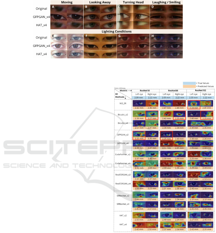
Figure 4: Challenges in estimating pupil diameter without and with SR: Participants A, B, C show head movements and gaze
shifts; Participant D shows eye size variation while smiling; Participants E, F, G, H experience different lighting effects—E
in bright light, F with a yellow tint, G’s face appearing red, and H’s face appearing blue.
bining SR with image-to-image translation models
like Pix2Net (Jin et al., 2024), which converts RGB
images to near-infrared (NIR), could improve fea-
ture extraction, particularly in low-contrast scenarios
where darker irises make pupil features difficult to
detect. Additionally, real-time SR techniques, such
as those introduced by (Zhan et al., 2021) and (Shi
et al., 2016), could enable mobile and web-based ap-
plications for real-time pupilometry without special-
ized equipment. These advancements will not only
enhance the accuracy of eye-tracking technologies but
also make them more accessible, laying a strong foun-
dation for future innovations in both pupilometry and
eye-tracking technology.
8 CONCLUSION
In this work, we investigated the role of SR tech-
niques in enhancing the accuracy of pupil diame-
ter prediction from webcam-based images, which is
crucial for assessing psychological and physiolog-
ical states. Our experiments, across multiple up-
scaling methods and neural network architectures,
demonstrate that SR can significantly refine the fea-
ture details necessary for more precise pupil mea-
surements. Key findings indicate that while the ben-
efits of SR are clear, they are not uniformly dis-
tributed across different scales and methods. For in-
stance, although traditional bicubic upscaling often
performs well, advanced SR techniques like Real-
ESRGAN and SRResNet generally provide superior
error rates under specific conditions. In conclusion,
while SR presents a promising avenue for enhancing
low-quality, webcam-derived images for pupilometry,
it requires nuanced application and thorough valida-
tion to fully realize its benefits.
Figure 5: Class Activation Map (Zhou et al., 2016) vi-
sualizations for the final convolutional layer of ResNet18,
ResNet50, and ResNet152 are shown for a test participant
viewing the same display color with No-SR, SRx2, and
SRx4 eye images. The true and predicted values represent
the original and estimated pupil diameters.
ACKNOWLEDGEMENTS
This work was supported by the DFG Interna-
tional Call on Artificial Intelligence “Learning Cy-
ICAART 2025 - 17th International Conference on Agents and Artificial Intelligence
382

clotron” (442581111) and the BMBF project Sus-
tainML (Grant 101070408).
REFERENCES
Agustsson, E. and Timofte, R. (2017). Ntire 2017 challenge
on single image super-resolution: Dataset and study.
In CVPRW, pages 126–135.
Apple Inc. (2024). Apple announces new accessibility fea-
tures, including eye tracking, music haptics, and vocal
shortcuts. Accessed: 2024-06-06.
Bashir, S. M. A., Wang, Y., Khan, M., and Niu, Y. (2021).
A comprehensive review of deep learning-based sin-
gle image super-resolution. PeerJ Computer Science,
7:e621.
Bhatt, A., Watanabe, K., Dengel, A., and Ishimaru, S.
(2024). Appearance-based gaze estimation with deep
neural networks: From data collection to evaluation.
International Journal of Activity and Behavior Com-
puting, 2024(1):1–15.
Caya, M. V. C., Jorel P. Rapisura, C., and Despabiladeras,
R. R. B. (2022). Development of pupil diameter
determination using tiny-yolo algorithm. In 2022
IEEE 14th International Conference on Humanoid,
Nanotechnology, Information Technology, Communi-
cation and Control, Environment, and Management
(HNICEM), pages 1–6.
Chen, C.-C., Chen, W.-H., Chiang, J.-S., Chien, C.-T., and
Chang, T. (2023a). Semantic segmentation using su-
per resolution technique as pre-processing. In IET In-
ternational Conference on Engineering Technologies
and Applications (ICETA 2023), volume 2023, pages
109–110. IET.
Chen, J., Jia, L., Zhang, J., Feng, Y., Zhao, X., and Tao, R.
(2024). Super-resolution for land surface temperature
retrieval images via cross-scale diffusion model using
reference images. Remote Sensing, 16(8):1356.
Chen, X., Wang, X., Zhou, J., Qiao, Y., and Dong, C.
(2023b). Activating more pixels in image super-
resolution transformer. In CVPR, pages 22367–22377.
Dembinsky, D., Watanabe, K., Dengel, A., and Ishimaru, S.
(2024a). Eye movement in a controlled dialogue set-
ting. In Proceedings of the 2024 Symposium on Eye
Tracking Research and Applications, ETRA ’24, New
York, NY, USA. Association for Computing Machin-
ery.
Dembinsky, D., Watanabe, K., Dengel, A., and Ishimaru,
S. (2024b). Gaze generation for avatars using gans.
IEEE Access, 12:101536–101548.
Dosovitskiy, A., Beyer, L., Kolesnikov, A., Weissenborn,
D., Zhai, X., Unterthiner, T., Dehghani, M., Minderer,
M., Heigold, G., Gelly, S., et al. (2020). An image is
worth 16x16 words: Transformers for image recogni-
tion at scale. arXiv preprint arXiv:2010.11929.
Elman, J. A., Panizzon, M. S., Hagler Jr, D. J., Eyler, L. T.,
Granholm, E. L., Fennema-Notestine, C., Lyons,
M. J., McEvoy, L. K., Franz, C. E., Dale, A. M., et al.
(2017). Task-evoked pupil dilation and bold variance
as indicators of locus coeruleus dysfunction. Cortex,
97:60–69.
Fathi, A. and Abdali-Mohammadi, F. (2015). Camera-based
eye blinks pattern detection for intelligent mouse. Sig-
nal, Image And Video Processing, 9:1907–1916.
Granholm, E. L., Panizzon, M. S., Elman, J. A., Jak, A. J.,
Hauger, R. L., Bondi, M. W., Lyons, M. J., Franz,
C. E., and Kremen, W. S. (2017). Pupillary responses
as a biomarker of early risk for alzheimer’s disease.
Journal of Alzheimer’s disease, 56(4):1419–1428.
Greinacher, R. and Voigt-Antons, J.-N. (2020). Accu-
racy assessment of arkit 2 based gaze estimation.
In Kurosu, M., editor, Human-Computer Interaction.
Design and User Experience, pages 439–449, Cham.
Springer International Publishing.
He, C., Xu, Y., Wu, Z., and Wei, Z. (2024). Connecting low-
level and high-level visions: A joint optimization for
hyperspectral image super-resolution and target detec-
tion. IEEE Transactions on Geoscience and Remote
Sensing.
He, K., Zhang, X., Ren, S., and Sun, J. (2016). Deep resid-
ual learning for image recognition. In CVPR, pages
770–778.
Hisadome, Y., Wu, T., Qin, J., and Sugano, Y. (2024).
Rotation-constrained cross-view feature fusion for
multi-view appearance-based gaze estimation. In
WACV, pages 5985–5994.
Hong, J., Shin, J., Choi, J., and Ko, M. (2024). Robust
eye blink detection using dual embedding video vision
transformer. In WACV, pages 6374–6384.
Huang, H., He, R., Sun, Z., Tan, T., et al. (2018). In-
trovae: Introspective variational autoencoders for pho-
tographic image synthesis. Advances in neural infor-
mation processing systems, 31.
Jiang, T., Yu, Q., Zhong, Y., and Shao, M. (2024). Plantsr:
Super-resolution improves object detection in plant
images. Journal of Imaging, 10(6):137.
Jin, Y., Park, I., Song, H., Ju, H., Nalcakan, Y., and Kim, S.
(2024). Pix2next: Leveraging vision foundation mod-
els for rgb to nir image translation. arXiv preprint
arXiv:2409.16706.
Joshi, S., Li, Y., Kalwani, R. M., and Gold, J. I. (2016). Re-
lationships between pupil diameter and neuronal ac-
tivity in the locus coeruleus, colliculi, and cingulate
cortex. Neuron, 89(1):221–234.
Kahneman, D. and Beatty, J. (1966). Pupil diameter and
load on memory. Science, 154(3756):1583–1585.
Karras, T., Laine, S., and Aila, T. (2018). A style-
based generator architecture for generative adver-
sarial networks. arxiv e-prints. arXiv preprint
arXiv:1812.04948.
Khokhlov, I., Davydenko, E., Osokin, I., Ryakin, I., Babaev,
A., Litvinenko, V., and Gorbachev, R. (2020). Tiny-
yolo object detection supplemented with geometrical
data. In 2020 IEEE 91st Vehicular Technology Con-
ference (VTC2020-Spring), pages 1–5. IEEE.
Kim, J., Oh, J., and Lee, K. M. (2024). Beyond image super-
resolution for image recognition with task-driven per-
ceptual loss. In CVPR, pages 2651–2661.
Webcam-Based Pupil Diameter Prediction Benefits from Upscaling
383

Kotani, J., Nakao, H., Yamada, I., Miyawaki, A., Mambo,
N., and Ono, Y. (2021). A novel method for mea-
suring the pupil diameter and pupillary light reflex of
healthy volunteers and patients with intracranial le-
sions using a newly developed pupilometer. Frontiers
in Medicine, 8.
Krejtz, K., Duchowski, A. T., Niedzielska, A., Biele, C.,
and Krejtz, I. (2018). Eye tracking cognitive load us-
ing pupil diameter and microsaccades with fixed gaze.
PloS one, 13(9):e0203629.
Kremen, W. S., Panizzon, M. S., Elman, J. A., Granholm,
E. L., Andreassen, O. A., Dale, A. M., Gillespie,
N. A., Gustavson, D. E., Logue, M. W., Lyons, M. J.,
et al. (2019). Pupillary dilation responses as a midlife
indicator of risk for alzheimer’s disease: association
with alzheimer’s disease polygenic risk. Neurobiol-
ogy of Aging, 83:114–121.
Kucewicz, M. T., Dolezal, J., Kremen, V., Berry, B. M.,
Miller, L. R., Magee, A. L., Fabian, V., and Worrell,
G. A. (2018). Pupil size reflects successful encoding
and recall of memory in humans. Scientific reports,
8(1):4949.
Ledig, C., Theis, L., Husz
´
ar, F., Caballero, J., Cunningham,
A., Acosta, A., Aitken, A., Tejani, A., Totz, J., Wang,
Z., et al. (2017). Photo-realistic single image super-
resolution using a generative adversarial network. In
CVPR, pages 4681–4690.
Li, X., Ren, Y., Jin, X., Lan, C., Wang, X., Zeng, W.,
Wang, X., and Chen, Z. (2023). Diffusion models for
image restoration and enhancement–a comprehensive
survey. arXiv preprint arXiv:2308.09388.
Liu, J. (2024). Improving image stitching effect using
super-resolution technique. International Journal of
Advanced Computer Science & Applications, 15(6).
Liu, M., Bian, S., and Lukowicz, P. (2022). Non-contact,
real-time eye blink detection with capacitive sensing.
In Proceedings of the 2022 ACM International Sympo-
sium on Wearable Computers, ISWC ’22, page 49–53,
New York, NY, USA. Association for Computing Ma-
chinery.
L
¨
udtke, H., Wilhelm, B., Adler, M., Schaeffel, F., and Wil-
helm, H. (1998). Mathematical procedures in data
recording and processing of pupillary fatigue waves.
Vision research, 38(19):2889–2896.
Micieli, G., Tassorelli, C., Martignoni, E., Pacchetti, C.,
Bruggi, P., Magri, M., and Nappi, G. (1991). Disor-
dered pupil reactivity in parkinson’s disease. Clinical
Autonomic Research, 1:55–58.
Moser, B. B., Raue, F., Frolov, S., Palacio, S., Hees, J.,
and Dengel, A. (2023). Hitchhiker’s guide to super-
resolution: Introduction and recent advances. IEEE
TPAMI, 45(8):9862–9882.
Moser, B. B., Raue, F., Palacio, S., Frolov, S., and Dengel,
A. (2024a). Latent dataset distillation with diffusion
models. arXiv preprint arXiv:2403.03881.
Moser, B. B., Shanbhag, A. S., Raue, F., Frolov, S., Palacio,
S., and Dengel, A. (2024b). Diffusion models, im-
age super-resolution and everything: A survey. arXiv
preprint arXiv:2401.00736.
Murillo, R., Crucilla, C., Schmittner, J., Hotchkiss, E., and
Pickworth, W. B. (2004). Pupillometry in the detec-
tion of concomitant drug use in opioid-maintained pa-
tients. Methods and findings in experimental and clin-
ical pharmacology, 26(4):271–275.
Murphy, P. R., O’connell, R. G., O’sullivan, M., Robert-
son, I. H., and Balsters, J. H. (2014). Pupil diameter
covaries with bold activity in human locus coeruleus.
Human brain mapping, 35(8):4140–4154.
Mustafa, A., Khan, S. H., Hayat, M., Shen, J., and Shao, L.
(2019). Image super-resolution as a defense against
adversarial attacks. arXiv preprint arXiv:1901.01677.
Na, B. and Fox, G. C. (2020). Object classifications
by image super-resolution preprocessing for convolu-
tional neural networks. Advances in Science, Tech-
nology and Engineering Systems Journal (ASTESJ),
5(2):476–483.
Ni, Y. and Sun, B. (2019). A remote free-head pupillometry
based on deep learning and binocular system. IEEE
Sensors Journal, 19(6):2362–2369.
O’Shea, G. and Komeili, M. (2023). Toward super-
resolution for appearance-based gaze estimation.
arXiv preprint arXiv:2303.10151.
Pedrotti, M., Mirzaei, M. A., Tedesco, A., Chardonnet,
J.-R., M
´
erienne, F., Benedetto, S., and Baccino, T.
(2014). Automatic stress classification with pupil di-
ameter analysis. International Journal of Human-
Computer Interaction, 30(3):220–236.
Pfleging, B., Fekety, D. K., Schmidt, A., and Kun, A. L.
(2016). A model relating pupil diameter to mental
workload and lighting conditions. In Proceedings of
the 2016 CHI Conference on Human Factors in Com-
puting Systems, CHI ’16, page 5776–5788, New York,
NY, USA. Association for Computing Machinery.
Reddy, L. F., Reavis, E. A., Wynn, J. K., and Green, M. F.
(2018). Pupillary responses to a cognitive effort task
in schizophrenia. Schizophrenia Research, 199:53–
57.
Sari, J. N., Hanung, A. N., Lukito, E. N., Santosa, P. I.,
and Ferdiana, R. (2016). A study on algorithms
of pupil diameter measurement. In 2016 2nd In-
ternational Conference on Science and Technology-
Computer (ICST), pages 188–193.
Shah, V., Watanabe, K., Moser, B. B., and Dengel, A.
(2024). Eyedentify: A dataset for pupil diameter es-
timation based on webcam images. arXiv preprint
arXiv:2407.11204.
Shi, W., Caballero, J., Husz
´
ar, F., Totz, J., Aitken, A. P.,
Bishop, R., Rueckert, D., and Wang, Z. (2016). Real-
time single image and video super-resolution using an
efficient sub-pixel convolutional neural network. In
CVPR, pages 1874–1883.
Song, L., Wang, Q., Liu, T., Li, H., Fan, J., Yang, J., and Hu,
B. (2022). Deep robust residual network for super-
resolution of 2d fetal brain mri. Scientific reports,
12(1):406.
Tales, A., Troscianko, T., Lush, D., Haworth, J., Wilcock,
G., and Butler, S. (2001). The pupillary light reflex in
aging and alzheimer’s disease. Aging (Milan, Italy),
13(6):473–478.
ICAART 2025 - 17th International Conference on Agents and Artificial Intelligence
384

Tang, J., Zhang, J., Chen, D., Al-Nabhan, N., and Huang,
C. (2021). Single-frame super-resolution for remote
sensing images based on improved deep recursive
residual network. EURASIP Journal on Image and
Video Processing, 2021:1–19.
Timofte, R., Agustsson, E., Van Gool, L., Yang, M.-H.,
and Zhang, L. (2017). Ntire 2017 challenge on sin-
gle image super-resolution: Methods and results. In
CVPRW, pages 114–125.
Van Den Brink, R. L., Murphy, P. R., and Nieuwenhuis, S.
(2016). Pupil diameter tracks lapses of attention. PloS
one, 11(10):e0165274.
Wang, X., Li, Y., Zhang, H., and Shan, Y. (2021a). Towards
real-world blind face restoration with generative facial
prior. In CVPR, pages 9168–9178.
Wang, X., Li, Y., Zhang, H., and Shan, Y. (2021b). Towards
real-world blind face restoration with generative facial
prior. In CVPR.
Wang, X., Xie, L., Dong, C., and Shan, Y. (2022). Realesr-
gan: Training real-world blind super-resolution with
pure synthetic data supplementary material. Computer
Vision Foundation open access, 1(2):2.
Yun, J.-S., Na, Y., Kim, H. H., Kim, H.-I., and Yoo, S. B.
(2022). Haze-net: High-frequency attentive super-
resolved gaze estimation in low-resolution face im-
ages. In ACCV, pages 3361–3378.
Zhan, Z., Gong, Y., Zhao, P., Yuan, G., Niu, W., Wu,
Y., Zhang, T., Jayaweera, M., Kaeli, D., Ren,
B., et al. (2021). Achieving on-mobile real-time
super-resolution with neural architecture and pruning
search. In ICCV, pages 4821–4831.
Zhang, Z. (1999). Flexible camera calibration by viewing a
plane from unknown orientations. In Proceedings of
the Seventh IEEE International Conference on Com-
puter Vision, volume 1, pages 666–673 vol.1.
Zhou, B., Khosla, A., Lapedriza, A., Oliva, A., and Tor-
ralba, A. (2016). Learning deep features for discrimi-
native localization. In CVPR, pages 2921–2929.
Zhou, S., Chan, K., Li, C., and Loy, C. C. (2022). Towards
robust blind face restoration with codebook lookup
transformer. NeurIPS, 35:30599–30611.
Webcam-Based Pupil Diameter Prediction Benefits from Upscaling
385
