
The Role of Self-Focusing During the Laser Microstructuring in the
Volume of Fused Silica
Anna V. Bogatskaya
1,2 a
, Ekaterina A. Volkova
3b
and Alexander M. Popov
1,2 c
1
Department of Physics, Lomonosov Moscow State University, 119991, Moscow, Russia
2
Lebedev Physical Institute, Russian Academy of Sciences, 119991, Moscow, Russia
3
Skobeltsyn Institute of Nuclear Physics, Lomonosov Moscow State University, 119991, Moscow, Russia
Keywords: Laser Microstructuring in Dielectrics, Birefringent Nanolattices, Fused Silica,
Multiphoton Ionization of Dielectrics, Plasma Formation, Numerical Modelling, Wave Equation.
Abstract: In this study we perform 3D self-consistent numerical simulations of a focused laser pulse exposure in the
bulk of fused silica. The model combines the second-order wave equation in cylindrical coordinates with a
rate equation for the density of charge carriers in the conduction band. Our results indicate that a dense plasma
formation near the focal plane effectively scatters and reflects the laser pulse. The coherent interference
between the incident and scattered laser waves creates regions of intense field ionization, resulting in periodic
plasma nanostructures along both the ρ- and z-axes. We also examine the impact of nonlinear refractive index
effects, which lead to pulse self-focusing. We should note that similar subwavelength, divergent structures in
material modification regions have been observed in recent experiments conducted under comparable laser
focusing conditions.
1 INTRODUCTION
In recent years, substantial research has been
dedicated to investigating the complex, multi-level
processes that modify the physical characteristics of
materials subjected to tightly focused femtosecond
laser pulses (Gattass and Mazur, 2008; Taylor et al.,
2007; Bulgakova et al., 2015). State-of-the-art
ultrafast laser systems have revealed new
mechanisms in how electromagnetic fields, plasma,
and materials interact. These interactions drive
various structural changes in transparent dielectric
materials, such as the formation of micro- and
nanoscale voids, densification zones, micro-tracks
(Shimotsuma et al., 2005; Sun et al., 2007; Beresna et
al., 2011; Dai et al., 2016; Mizeikis et al., 2009), as
well as periodic refractive index shifts (Schaffer et al.,
2001; Wang et al., 2007; Mermillod-Blondin et al.,
2008) and other effects.
One of the most notable achievements has been
the development of periodic subwavelength
structures with high optical contrast between
a
https://orcid.org/0000-0002-1538-3433
b
https://orcid.org/0000-0002-4883-3349
c
https://orcid.org/0000-0002-7300-3785
modified zones, which are particularly promising for
applications in optical memory, micro-photonic
crystals, optical couplers, binary storage, and other
fields (Musgraves et al., 2011; Tan et al., 2016).
Among these materials, fused silica glass has become
especially important since the groundbreaking work
of (Shimotsuma et al. 2003), which first introduced
the concept of birefringent volume nanogratings in
this medium. Subsequent research by various groups
(Desmarchelier et al., 2015; Bulgakova et al., 2013)
has highlighted key mechanisms involved in creating
these structures. These mechanisms include coupling
between electron plasma waves and incident light
(Shimotsuma et al., 2005; Shimotsuma et al., 2003),
the formation of nanoplasmas due to field localization
and their self-organization into nanoscale patterns
(Bhardwaj et al., 2006; Taylor et al., 2008), and the
confinement and clustering of exciton-polaritons
(Beresna et al., 2012). However, the lack of
comprehensive theoretical models to confirm each
proposed mechanism, as well as an incomplete
understanding of the necessary conditions, still
120
Bogatskaya, A. V., Volkova, E. A. and Popov, A. M.
The Role of Self-Focusing During the Laser Microstructuring in the Volume of Fused Silica.
DOI: 10.5220/0013248900003902
In Proceedings of the 13th International Conference on Photonics, Optics and Laser Technology (PHOTOPTICS 2025), pages 120-125
ISBN: 978-989-758-736-8; ISSN: 2184-4364
Copyright © 2025 by Paper published under CC license (CC BY-NC-ND 4.0)

presents challenges to the controlled and precise laser
fabrication of these complex volume nanopatterns.
In this work, we carry out fully self-consistent 3D
numerical simulations of the propagation of an
intense, tightly focused Yb-doped fiber laser pulse
( 𝜆
=1030 nm) in fused silica, alongside the
dynamics of laser-induced solid-state plasma. The
simulations explore a range of pulse durations, peak
intensities, and focusing parameters. By including the
curvature of the incident laser pulse front and
considering scattering and diffraction effects in
cylindrical geometry, we identified a new mechanism
of plasma self-organization in conditions of tight laser
beam focusing. The findings reveal that dense plasma
formation near the focal area effectively scatters the
laser wave. Due to the interference between the
incident plane wave and the reflected, highly non-
planar wave in the pre-focal area, a detailed pattern of
laser field amplitude peaks and troughs emerges,
leading to a structured subwavelength plasma
distribution. It is important to note that such
mechanism of laser nanostructuring has not been
mentioned in previous studies, which certainly opens
up prospects for further progress in the area of
controlled laser writing in the volume of transparent
dielectrics. We also examine the effect of refractive
index nonlinearity which can lead to partial self-
focusing compensating the pulse defocusing by
plasma electrons.
2 MODELLING AND METHODS
Our modelling involves solving the second-order
wave equation for a focused linearly polarized laser
pulse moving through the fused silica in a cylindrical
geometry:
𝜌
+
=
с
+
. (1)
In this equation, 𝐸=𝐸(𝜌,𝑧,𝑡) denotes the electric
field strength of the laser pulse as it propagates in the
z-direction, 𝑛 is the fused silica refractive index
which includes both linear and nonlinear terms: 𝑛=
𝑛
+𝑛
𝐼. Here 𝑛
=1.45 is the linear part of the
refractive index, while the second term being
proportional to laser intensity 𝐼
(
𝜌,𝑧,𝑡
)
stands for the
cubic nonlinearity. According to (Vermeulen et al,
2023) we put 𝑛
=2.23×10
cm
2
/W. The second
term 𝑗(𝜌,𝑧,𝑡) in right part of equation (2) is the field
induced electron current. This current includes
contributions from both the polarization of charge
carriers within the conduction band and ionization
effects caused by transitions from the valence band to
the conduction band. Both of these currents depend
on the evolution of electron density 𝑛
(𝜌,𝑧,𝑡) within
the conduction band, governed by the following
equation:
𝜕𝑛
𝜕𝑡
=𝐷
1
𝜌
𝜕
𝜕𝜌
𝜌
𝜕𝑛
𝜕𝜌
+
𝜕
𝑛
𝜕𝑧
+𝑊
(
𝐼
)(
𝑁
−𝑛
)
+𝜈
(
𝐼
)
𝑛
1 −
𝑛
𝑁
−
𝑛
𝜏
(2)
Here 𝐷 ≈300 cm
2
/s is the electron diffusion
coefficient, 𝜏
=150 fs is the average electron
recombination time (Audebert, et. al., 1990), 𝑊
(
𝐼
)
and 𝜈
(𝐼) represent the rates of field-driven and
electron-impact ionization, 𝐼(𝜌,𝑧,𝑡) is the laser
radiation intensity, 𝑁
=2.1∙10
см
-3
is an atomic
density of fused silica. The laser intensity 𝐼
(
𝜌,𝑧,𝑡
)
is
derived from the electric field as follows: 𝐼
(
𝜌,𝑧,𝑡
)
=
〈
𝑛
𝐸
(
𝜌,𝑧,𝑡
)〉
, where brackets mean averaging
over the period of wave field oscillations. To
calculate the ionization probability we utilize the
general Keldysh formula (Keldysh, 1964;
Bogatskaya, et. al., 2023).
To account for the energy losses of the laser pulse
due to the field ionization process, we introduce an
ionization current 𝑗
(𝑧,𝑡) on the right side of Eq.
(1). This current can be expressed as
𝑗
(
𝜌,𝑧,𝑡
)
=
〈
〉
=
(3)
=
〈
〉
𝑊
𝐼
(
𝜌,𝑧,𝑡
)
(𝑁
−𝑛
(
𝜌,𝑧,𝑡
)
),
where 𝜕𝑛
𝜕𝑡
⁄
is the rate of electron density
production due to field ionization,
〈
𝐸
〉
is an averaged
over the period field
|
𝐸
(
𝜌,𝑧,𝑡
)|
. One should also
consider the polarization current 𝑗
(𝜌,𝑧,𝑡) of
electrons in the conduction band, which is induced by
the laser pulse. This current can be described via the
Drude model:
+𝜈
𝑗
=
∗
𝐸
(
𝜌,𝑧,𝑡
)
. (4)
Here 𝜈
≈2×10
s
-1
is the transport
collisional frequency. Total current in (1) reads
𝑗=𝑗
+𝑗
. (5)
The electron impact ionization is also included in
the right part of equation (2). The frequency of
electron impact ionization is expressed in terms
[Raizer. 1977]:
𝜈
(𝐼)=
(
,,
)
∗
. (6)
The Role of Self-Focusing During the Laser Microstructuring in the Volume of Fused Silica
121
Here 𝑚
∗
=0.5𝑚
is the effective mass of charge
carriers in fused silica, 𝜔 ≈1.83× 10
s
-1
is the
laser frequency corresponding to 𝜆
=1030 nm.
Our simulations use laser pulses with Gaussian
radial profiles at the focal plane 𝑧=0
( 𝐸~𝑒𝑥𝑝
(
−(𝜌 𝜌
⁄
)
)
and sin-squared temporal
envelope (𝐸~𝐸
𝑠𝑖𝑛
,𝑡∈(0,𝜏
)). To obtain
such pulse, the wave equation similar to (1) but
without the term of the electric current in the right-
hand part was integrated with zero initial conditions
for electric field strength and its derivative over time
and the boundary condition at 𝑧=0:
𝜕𝐸
(
𝜌,𝑧,𝑡
)
𝜕𝑧
=
𝜔𝑛
𝑐
𝐸
exp
(
−
(
𝜌𝜌
⁄)
)
𝑠𝑖𝑛
𝜋𝑡
𝜏
cos
(
𝜔𝑡
)
,
𝑡∈0
∗
,𝜏
(7)
𝜕𝐸
(
𝜌,𝑧= 0,𝑡
)
𝜕𝑧
=0, 𝑡>𝜏
The obtained pulse is considered to be the initial
one in further simulations. If one reverses the time
scale, this pulse will move back in negative direction
towards the focal plane 𝑧=0.
The focal spot radius varies from 𝜌
=1.7 to 3
mkm, while the pulse duration ranges from 𝜏
=12.5
to 75 fs. These parameters correspond to pulse
focusing within the fused silica volume with no initial
free charge carriers. The spatial pulse length falls
within ℓ
=
(
𝑐𝑛
⁄)
𝜏
~2.5 − 15 mkm. For pulses
with a 50 fs duration and focal radius 𝜌
=2.5 mkm
the energy range was set to 0.024 – 0.34 mkJ, yielding
peak intensities of approximately ~5 × 10
−7×
10
W/cm
2
at the focal plane (here we again neglect
the birth of electron density in the focal plane region
leading to the defocusing as a consequence, a decrease
in focal intensity). For considered pulse parameters, the
rate of field ionization dominates in comparison with
electron impact ionization (Bogatskaya et al, 2023).
Therefore, the electron avalanche, commonly seen in
breakdown processes in gases and solids over
microsecond and nanosecond timescales, does not
control the plasma dynamics in this case. The
equations (1) and (2) were solved jointly within the
spatial domain
𝜌,𝑧
=
0−𝜌
,0−𝐿
with 𝜌
=
30 mkm and, 𝐿=120 mkm. Initial pulse (𝑡=0) was
centered around 𝑧
≈105 µm and propagated in the
negative z-direction, achieving peak focus close to the
origin ( 𝑧=30 mkm). The numerical integration
approach for the second-order wave equation is briefly
described in (Bogatskaya et al, 2019).
3 RESULTS AND DISCUSSION
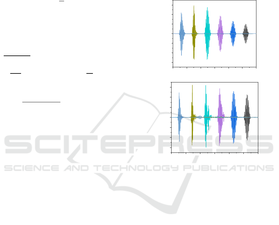
We start our analysis with the data on pulse focusing
during the propagation in the bulk of fused silica. In
Fig.1 we perform the simulations of on-axis field
distributions for two values of laser peak intensity.
0 20 40 60 80 100 120
-1.0
-0.7
-0.3
0.0
0.3
0.7
1.0
E(z,t,
1
=0), arb.un.
z, mkm
a
020406080100120
-1.0
-0.7
-0.3
0.0
0.3
0.7
1.0
E(z,t,
−
=0), arb.un.
z
, mkm
b
Figure 1: On-axis electric field distributions for different
instants of time (intervals between time points ~ 0.1 ps).
The pulse moves from right to left and focuses at point 𝑧=
30 mkm. Pulse peak intensity is 10
W/cm
2
(a), 5·10
W/cm
2
(b). Focal spot radius is 𝜌
=2.5 mkm, pulse
duration is 50 fs.
One can see that for more intense laser pulse
strong defocusing takes place due to the formation of
dense plasma near the focal area. Indeed, the
distributions of electron concentration for the
intensity 5·10
W/cm
2
(see Fig.2) indicate the
values of electron density more than 10
cm
-3
. In
this case plasma electrons significantly defocuses and
reflects laser radiation, which results in appearing
rather regular structures both in the 𝜌- and z-axis
direction. The formation of such patterns can be
attributed to the coherent interference between a
nearly planar incident wave in the pre-focal region
and a significantly non-planar (almost spherical)
wave scattered by a dense plasma burst [Bogatskaya,
et al, 2024].
PHOTOPTICS 2025 - 13th International Conference on Photonics, Optics and Laser Technology
122
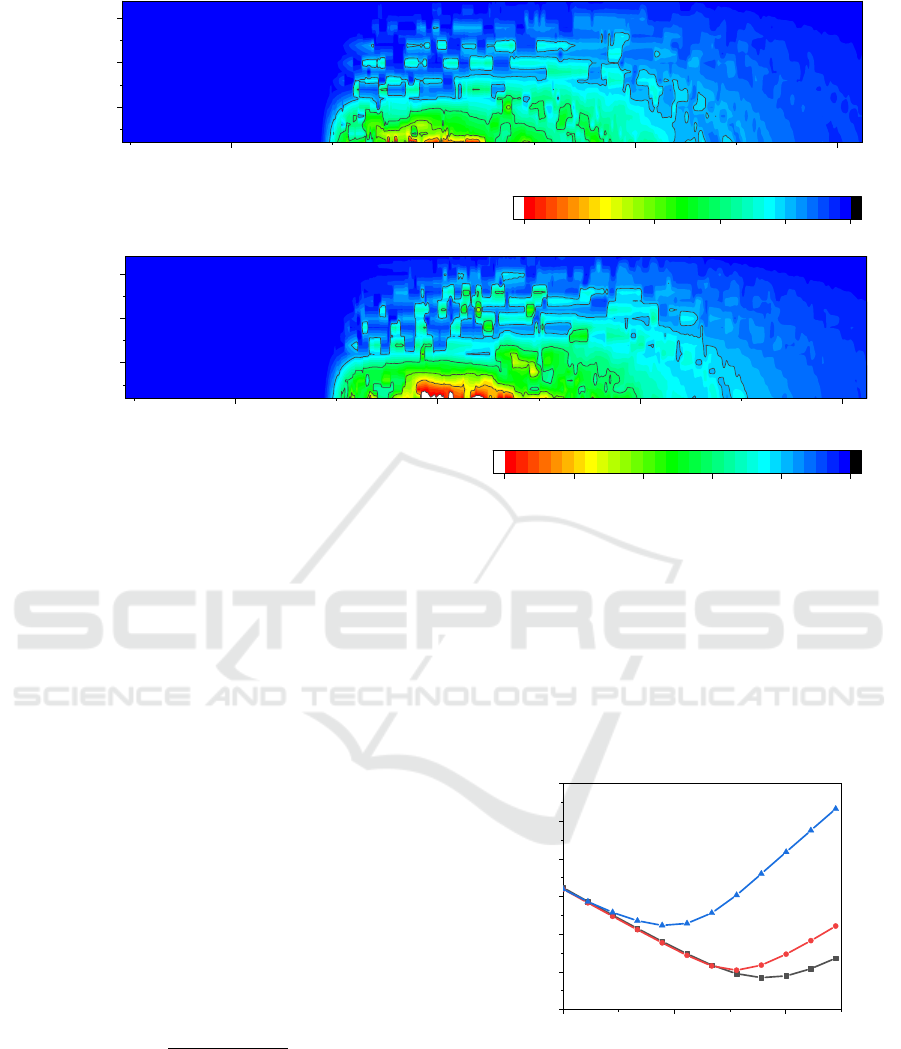
Figure 2: Profiles of electron density in the volume of fused silica formed by the 50-fs laser pulse with the peak intensity
5·10^13 W/cm2 (pulse energy is 0.24 mkJ). Graph (a) represents data in the absence of self-focusing effect, (b) – accounting
the self-focusing effect. Focal spot radius is ρ_0=2.5 mkm, the position of focal plane is 30 mkm.
In Fig.2 we also analyze the effect of refractive index
nonlinearity on the plasma microstructures formation.
In particular, Fig 2b presents simulations accounting
𝑛
. Simulations have shown that in the absence of
electrons the threshold of self-focusing in fused silica
is about 6 MW. However, due to the strong
defocusing of plasma electrons, the presence of self-
focusing induced by the nonlinear refractive index
has little effect on the overall picture of plasma
formations. Thus, in Figure 2b one can observe that
considering nonlinearity leads to a slight increase in
the electron concentration in the paraxial zone near
the focus.
The effect of electrons on the laser beam focusing
can be clearly observed in Figure 3, which shows the
dependence of the mean radius of the laser pulse
during its propagation for different pulse intensities.
We calculate this radius, using the following formula:
〈
𝜌(𝑡)
〉
=
(,,)
(,,)
. (7)
At the initial instant of time the pulse is located at the
point 𝑧=105 mkm and moves from right to left
towards the focal plane at 𝑧=30 mkm. Curve 1
corresponds to the low energy pulse propagation in
the absence of ionization. It can be seen that an
increase of pulse energy leads to an increase in the
observed beam size and a shift in the position the
focal plane (minimum value of
〈
𝜌
〉
). An increase in
the focal spot size leads to a decrease in the peak
intensity of the beam, as a result, ionization saturation
occurs in the sample volume. This effect has been
repeatedly mentioned in a number of works on
focused radiation exposure in solid dielectrics
(Zheltikov, 2009; Rudenko, et al, 2023).
0.0
2.0x10
-13
4.0x10
-13
0
2
4
6
8
10
12
2
3
<ρ>, mkm
propagation time, s
1
Figure 3: The radial size of the pulse
〈
𝜌
〉
during its
propagation for different pulse intensities: 1 – weak pulse,
no plasma formation, 2 - 10
W/cm
2
, 3 - 6·10
W/cm
2
.
Plasma-free focal spot radius is 𝜌
=2.5 mkm, pulse
duration is 50 fs.
This study focuses on the consideration of linear
polarization of the laser pulse, however, the influence
of polarization on the femtosecond laser writing
20 40 60 80
0.00025
0.00056
0.00088
z, mkm
ρ, mkm
0.0
3.0x10
19
6.0x10
19
9.0x10
19
1.2x10
20
1.5x10
20
n
e
, cm
-3
a
20 40 60 80
0.00025
0.00056
0.00088
z, mkm
ρ
, mkm
0.0
3.0x10
19
6.0x10
19
9.0x10
19
1.2x10
20
1.5x10
20
n
e
, cm
-3
b
The Role of Self-Focusing During the Laser Microstructuring in the Volume of Fused Silica
123

process has been investigated in several experimental
studies. For example, in (Lei et al, 2023) they showed
that, contrary to intuitive expectations, ultrafast laser
direct writing with elliptical polarization in silica
glass results in birefringence approximately twice as
large as that observed with linearly polarized light,
although nonlinear absorption in the case of elliptical
polarization is about 2.5 times weaker. Moreover, the
use of laser pulses with different polarizations allows
for the creation of more complex nanostructure
topologies. However, the lack of theoretical studies at
present complicates the possibility of controlled
polarization-dependent laser writing of
nanostructures.
4 CONCLUSIONS
We performed the numerical study of the effect of
dense plasma formation in the volume of fused silica
exposed by intense tightly focused femtosecond IR
laser pulse. It was demonstrated that the plasma
object with electron density at a level ~1 − 2 × 10
cm
-3
arises in the pre-focal plane under the conditions
of tight focusing. The formed plasma effectively
scatters the incident femtosecond laser pulse
producing the region of effective wave interference.
As a result, the spatial distribution of the electron
production rate is characterized by rather sharp
maxima located in the bunches of the standing wave.
These maxima lead to the formation of periodic
subwavelength regions of dense plasma both in ρ- and
z-directions. It is important to note that the identified
mechanism of volumetric self-organization is
associated with a strong curvature of the front of the
reflected from plasma laser wave. It was shown that
due to strong beam defocusing by plasma electrons
the effect of pulse self-focusing is negligible.
Importantly, that the obtained profiles of plasma
nanostructures are found to be in good agreement
with SEM images of nanomodifications inscribed in
bulk fused and crystal silica accumulation regime
under the multi-pulse exposure (Zhang et al, 2019;
Gulina et al, 2024). Thus, in the work (Zhang et al,
2019), the formation of periodicity in a quartz crystal
was studied under the exposure of tightly focused
laser pulses with wavelengths of 1030 and 800 nm at
different pulse energies. The results of the experiment
demonstrated the formation of extended quasi-
periodic nanostructures, the spatial dimensions of
which increase with increasing energy input. The
similarity of the formed structures with the structures
written in the volume of fused silica under the
relatively equal laser focusing conditions, given in the
work of (Gulina et al, 2024), indicates the similarity
of the mechanisms of the initial (plasma) stage of the
nanostructuring, but later stages associated with the
formation of various defects and melting zones may
depend on a specific material, which requires further
analysis.
In summary, the importance of understanding the
process of fabrication of such periodic nanopatterns
is determined by their extensive application across
various fields, including optical polarizing elements
and devices, light waveguides, micro-photonic
crystals, binary storage components, and more (Tan
et al., 2016).
ACKNOWLEDGEMENTS
This research was funded by the Russian Science
Foundation (project no. 22-72-10076).
REFERENCES
Audebert, P.; Daguzan, Ph.; Dos Santos, A.; Gauthier, J. C.;
Geindre, J. P.; Guizard, S.; Hamoniaux, G.; Krastev, K.;
Martin, P.; Petite, G.; and Antonetti, A. (1994). Phys.
Rev. Lett., 73 (14), 1990.
Beresna, M.; Gecevičius, M.; Bulgakova, N. M.; Kazansky,
P. G. (2011). Opt. Express, 19, 18989.
Beresna, M.; Gecevičius, M.; Kazansky, P. G.; Taylor, T.;
Kavokin, A. (2012). Appl. Phys. Lett., 101, 053120.
Bhardwaj, V. R.; Simova, E.; Rajeev, P. P.; Hnatovsky, C.;
Taylor, R. S.; Rayner, D. M.; Corkum, P.B. (2006).
Phys. Rev. Lett., 96, 057404.
Bogatskaya, A. V.; Volkova, E. A.; Popov, A. M., (2019).
Laser Phys., 29, 086002.
Bogatskaya, A.; Gulina, Yu.; Smirnov, N.; Gritsenko, I.;
Kudryashov, S.; Popov, A. (2023). Photonics, 10, 515.
Bogatskaya, A. V.; Volkova, E. A.; Popov, A. M., (2024).
EPL, 147 (3), 35001.
Bulgakova, N. M.; Zhukov, V. P.; Meshcheryakov, Yu. P.
(2013). Appl. Phys. B, 113(3), 437-449.
Bulgakova, N. M.; Zhukov, V. P.; Sonina, S. V.;
Meshcheryakov, Y.P. (2015). J. Appl. Phys., 118 (23),
233108.
Dai, Y.; Patel, A.; Song, J.; Beresna, M.; and Kazansky, P.
G. (2016). Opt. Express, 24, 19344
Desmarchelier, R.; Poumellec, B.; Brisset, F.; Mazerat, S.
and Lancry, M. (2015). World Journal of Nano Science
and Engineering, 5, 115-125.
Gattass, R. R. and Mazur, E. (2008). Nature Photonics, 2,
219-225.
Gulina, Yu.; Rupasov, A.; Krasin, G.; Busleev, N.;
Gritsenko, I.; Bogatskaya, A.; Kudryashov, S. (2024).
JETP Lett., 119 (9), 638 – 644.
Keldysh, L.V. (1964). JETP, 20, 1307–1314.
PHOTOPTICS 2025 - 13th International Conference on Photonics, Optics and Laser Technology
124

Kudryashov, S. I.; Danilov, P. A.; Smaev, M. P.; Rupasov,
A. E.; Zolot’ko, A. S.; Ionin A. A.; Zakoldaev, R. A.
(2021). JETP Lett., 113, 493-497.
Kudryashov, S.; Rupasov, A.; Kosobokov, M.;
Akhmatkhanov, A.; Krasin, G.; Danilov, P.; Lisjikh, B.;
Abramov, A.; Greshnyakov, E.; Kuzmin, E.; et al.
(2022). Nanomaterials, 12, 4303.
Kudryashov, S.; Rupasov, A.; Smayev, M.; Danilov, P.;
Kuzmin, E.; Mushkarina, I.; Gorevoy, A.; Bogatskaya,
A.; and Zolot’ko, A. (2023) Nanomaterials, 13(6),
1133.
Lei, Y., Shayeganrad, G., Wang, H. Sakakura, M., Yu, Y.,
Wang, L., Kliukin, D., Skuja, L., Svirko. Y., Kazansky,
P. (2023). Light Sci Appl, 12, 74.
Mazur, E. (2008). Nature Photon, 2, 219–225.
Mermillod-Blondin, A.; Burakov, I. M.; Meshcheryakov,
Y. P.; Bulgakova, N. M.; Audouard, E.; Rosenfeld, A.;
Husakou, A.; Hertel, I. V.; Stoian, R. (2008). Phys. Rev.
B, 77, 104205.
Mizeikis, V.; Juodkazis, S.; Balciunas, T.; Misawa, H.;
Kudryashov, S.I.; Ionin, A.A.; Zvorykin, V.D. (2009).
J. Appl. Phys., 105, 123106.
Musgraves, J.; Richardson, K.; Jain, H. (2011). Opt. Mater.
Express, 1, 921-935.
Raizer. Yu. P. (1977). Laser induced discharge phenomena
(Consultants Bureau, New York)
Rudenko, A.; Moloney, J. M.; and Polynkin, P. (2023).
Phys. Rev. Applied 20, 064035.
Schaffer, C. B.; Brodeur, A.; García, J. F.; Mazur, E.
(2001). Opt. Lett., 26, 93.
Shimotsuma, Y.; Kazansky, P. G.; Qiu, J. R.; Hirao, K.
(2003). Phys. Rev. Lett., 91, 247405.
Shimotsuma, Y.; Hirao, K.; Qiu, J. R.; Kazansky, P. G.
(2005). Modern Phys. Lett. B, 19, 225.
Sun, H.Y.; Song, J.; Li, C.B.; Xu, J.; Wang, X. S.; Cheng,
Y.; Xu, Z. Z.; Qiu, J. R.; Jia, T. (2007). Appl. Phys. A,
88, 285.
Tan, D.; Sharafudeen, K.N.; Yue, Y.; Qiu, J. (2016).
Progress in Materials Science, 76, 154-228.
Taylor, R. S., Hnatovsky, C., Simova, E., Pattathilet, R.
(2007). Optics Letters, 32 (19), 2888-2890.
Taylor, R.; Hnatovsky, C.; Simova, E. (2008). Laser
Photonics Rev., 2, 26.
Vermeulen, N.; Espinosa, D.; Ball, A. et al (2023).
J. Phys.
Photonics, 5, 035001.
Wang, Z.; Sugioka, K.; Hanada, Y.; Midorikawa, K.
(2007). Appl. Phys. A, 88, 699.
Zhang, F.; Nie, Z.; Huang, H.; Ma, L.; Tang, H.; Hao, M.;
Qiu, J. (2019), Opt. Express, 27, 6442-6450.
Zheltikov. A. M. (2009). JETP Lett., 90, 90–95.
The Role of Self-Focusing During the Laser Microstructuring in the Volume of Fused Silica
125
