
Optical Fiber Probe Based on Localized Surface Plasmon Resonance
of Gold Nanostructures for Chemical Sensing
Amin Moslemi
1
, Lucia Sansone
2
, Flavio Esposito
1 a
, Carlos Marques
3,4
,
Stefania Campopiano
1 b
, Michele Giordano
2
and Agostino Iadicicco
1 c
1
Department of Engineering, University of Naples “Parthenope”, 80143 Naples, Italy
2
Institute for Polymers, Composites, and Biomaterials, National Research Council of Italy, IPCB-CNR, 80055 Portici, Italy
3
CICECO – Aveiro Institute of Materials, Physics Department, University of Aveiro, Aveiro 3810-193, Portugal
4
Department of Physics, VSB – Technical University of Ostrava, Ostrava 70800, Czech Republic
Keywords: Optical Fiber Sensors, Localized Surface Plasmon Resonance, Plasmonics, Biosensors, Chemical Sensors.
Abstract: In this study, we present an experimental investigation of highly sensitive optical fiber sensors utilizing
localized surface plasmon resonance (LSPR), achieved by depositing gold nanoparticles (NP) onto uncladded
silica multi-mode fiber. This setup takes advantage of the unique optical characteristics of optical fiber sensors
and plasmonic resonance provided by gold NPs. The experimental results demonstrated a maximum
sensitivity of about 130 nm/RIU in water solution, for an LSPR wavelength at 560 nm. As a study case, the
sensor was used to detect Thiram, a common agricultural pesticide, exhibiting a wide detection range from
10 nM to 100 µM, with a significant wavelength shift up to 4 nm. Moreover, a preliminary study involving
the use of nanostar-based optical fiber sensors is comparatively provided. The highest sensitivity makes this
approach highly promising for a range of applications, including environmental monitoring, biomedical
diagnostics, and chemical detection.
1 INTRODUCTION
Localized surface plasmon resonance (LSPR) refers
to the coherent oscillation of surface conduction
electrons in noble metal nanoparticles induced by
electromagnetic radiation at the nanoscale (Ma et al.,
2021; Mayer & Hafner, 2011). It is widely utilized for
sensing applications, with the LSPR absorption band
and its spectral position highly dependent on the
electrical properties of the noble metal (typically gold
or silver), the size and shape of the nanostructures,
and the dielectric properties of the surrounding
medium (Yaghubi et al., 2020). Consequently, these
devices are emerging as advanced sensors for
chemical and biological detection due to their
exceptional sensitivity to variations in the
surrounding medium refractive index (SRI) (Jeon et
al., 2019; Ma et al., 2021).
While early investigations of LSPR focused on
spherical gold nanoparticles (NPs), researchers are
a
https://orcid.org/0000-0003-1187-5825
b
https://orcid.org/0000-0002-2987-9122
c
https://orcid.org/0000-0002-3540-7316
now exploring a variety of sizes, shapes, and
materials. Nanostars (NSs) and nanorods (NRs), for
example, have different LSPR attenuation bands
compared to spherical NPs due to variations in their
shape and structural properties. These variations
allow for the generation of multiple resonances and
the ability to tune both the resonance wavelength and
sensitivity (He et al., 2020; Ringe et al., 2010;
Shabaninezhad & Ramakrishna, 2019; Ueno et al.,
2007). As a result, nanostructures with increased
aspect ratio offer high sensitivity for label-free
biosensing and chemical detection (Yuan et al.,
2012). For instance, Nguyen et al. introduced a
sensitive nanoplasmonic biosensor capable of
detecting two key epigenetic biomarkers using NSs:
methyl-CpG and MBD2, with detection limits of one
5-methylcytosine molecule and 125 fM MBD2,
respectively (Nguyen et al., 2015). Dondapati et al.
reported streptavidin binding to biotin-modified NSs
causing a plasmon resonance shift of 2.3 nm at
Moslemi, A., Sansone, L., Esposito, F., Marques, C., Campopiano, S., Giordano, M. and Iadicicco, A.
Optical Fiber Probe Based on Localized Surface Plasmon Resonance of Gold Nanostructures for Chemical Sensing.
DOI: 10.5220/0013395000003902
Paper published under CC license (CC BY-NC-ND 4.0)
In Proceedings of the 13th International Conference on Photonics, Optics and Laser Technology (PHOTOPTICS 2025), pages 157-162
ISBN: 978-989-758-736-8; ISSN: 2184-4364
Proceedings Copyright © 2025 by SCITEPRESS – Science and Technology Publications, Lda.
157
concentrations as low as 0.1 nM. (Dondapati et al.,
2010). Finally, Hashemi et al. developed a highly
sensitive electrochemical nanosensor to detect
monoclonal IgG antibodies against the SARS-CoV-2
S1 protein in blood within 1 minute. The sensor,
utilizing activated graphene and gold NSs, achieved
an ultra-low detection limit of 0.18∙10⁻
19
%V/V
(Alireza Hashemi et al., 2021).
Fiber optic sensors are increasingly popular due to
their advantages, including high sensitivity, improved
signal-to-noise ratio, cost-effectiveness, and the
ability to operate in harsh environments. These
characteristics make them suitable for a wide range of
chemical and biological sensing applications
(Choudhary et al., 2025; Gandhi et al., 2019;
Ricciardi et al., 2015).
While there is extensive literature on using
spherical NPs with optical fiber sensors, the
integration of other types of nanostructures with
optical fibers (Cennamo et al., 2013; Xiao et al.,
2025), however, is less explored, with only a few
studies available. For example, Cennamo et al.
introduced a novel optical chemical sensor that uses
molecularly imprinted polymer and NSs on plastic
optical fibers for the selective detection of
Trinitrotoluene in aqueous solutions. The device
achieves a sensitivity of 8.3∙10⁵ nm/M, 30 times
higher than previous gold layer SPR sensors
(Cennamo et al., 2015). In another study, Dos Santos
et al. utilized high aspect-ratio gold-silver nanorods,
which demonstrated a refractive index sensitivity of
1720 nm/RIU at 1350 nm (O-band) and 2325 nm/RIU
at 1550 nm (L-band). Using a side-polished optical
fiber, glyphosate detection they achieved a detection
limit improvement from 724 to 85 mg/L by shifting
to the C/L bands (dos Santos et al., 2025).
In this work, fiber optic probe based on spherical
gold nanoparticles properly grafted onto etched
multi-mode fiber (MMF) surface are evaluated for
chemical sensing applications. Specifically, its
performance has been assessed through the detection
of a dangerous compound like Thiram pesticide.
Moreover, a preliminary comparative study on the
use of gold-silver NSs in place of gold NPs is also
presented.
2 FABRICATION OF GOLD
NANOPARTICLES AND THEIR
CHARACTERIZATION
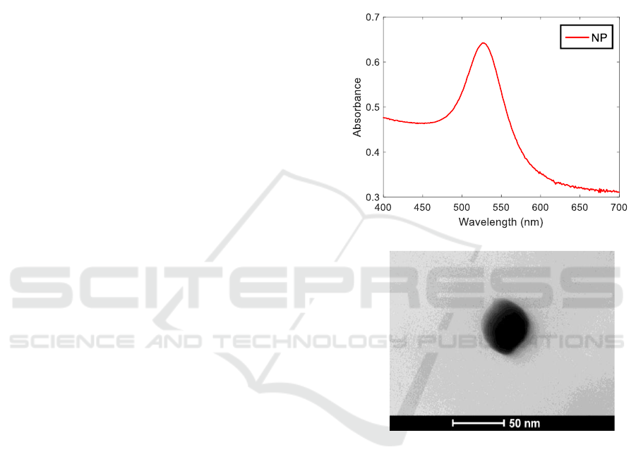
Gold NPs with spherical shapes were synthesized
using the well-assessed Turkevich method (Moslemi
et al., 2024). Figure 1(A) presents the UV-Vis spectra
recorded with an Agilent Cary 60 spectrophotometer
using quartz cuvettes, showing the results for
prepared NP samples. The absorbance peak centred at
531 nm is clearly visible. The morphological
characterization is provided by TEM images as in
Figure 1(B) where an average diameter of 40 nm can
be observed.
Figure 1: (A) UV-Vis spectra of gold NPs in solution; and
(B) TEM image of gold NP.
3 FABRICATION AND
CHARACTERIZATION OF THE
OPTICAL FIBER PROBE
3.1 Fabrication of the Probe
The basic sensor configuration is illustrated in Figure
2. It features an etched silica MMF with a mirrored
tip to enable reflected signal readout. The uncladded
etched region of the fiber is coated with metallic
nanostructures. The reflected signal from the probe
exhibits an attenuation band corresponding to the
excitation of LSPR.
(A)
(B)
AOMatDev 2025 - Special Session on Advanced Optical Materials and Devices
158
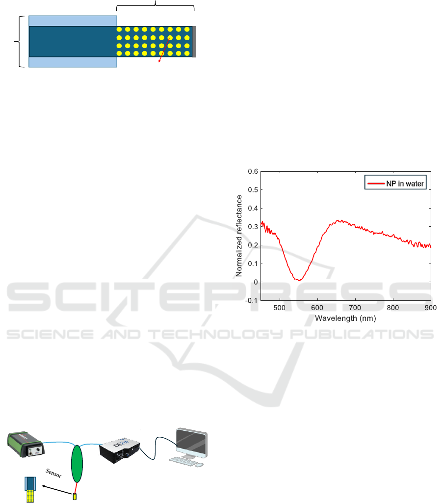
Figure 2: Schematic of the optical fiber probe.
For the fabrication, a few cm long piece of a
105/125 µm core/cladding MMF model Thorlabs
FG105LCA was etched for 25 minutes using 24%
hydrofluoric acid until the diameter was reduced to
95 μm, i.e. to remove the cladding and expose the
core. This allows the core modes to interact with
external media via the evanescent wave.
The silica surface of the etched fiber is then
silanized to facilitate the grafting of metallic
nanostructures. The process involves the following
steps. First, the fiber is immersed in piranha solution
for one hour to promote the formation of hydroxyl
groups, followed by thorough rinsing with water.
Next, it is immersed in an 5% w/w (3-
Aminopropyl)triethoxysilane (APTES) solution for
two hours to achieve surface silanization (Ben
Haddada et al., 2013). Afterward, the fiber is cleaned
with acetone and left to air-dry overnight.
Finally, to enable the reflection-based readout
approach, the fiber tip was cut and coated with a silver
layer to create a mirror.
Next, the functionalized fiber was immersed in the
metallic nanostructure solution prepared as detailed
in Section 2 for approximately four hours. The fiber
was then removed from the solution, left to dry
overnight, and washed five times with water to
remove loosely attached nanostructures.
Figure 3: Schematic of the optoelectronic readout setup.
An optoelectronic readout setup is necessary to
monitor the reflection spectrum of the so-prepared
sensing probe. The schematic of the setup is shown in
Figure 3: it consists of a broadband light source
(Avantes AvaLight-HAL-S-Mini) to provide incident
light. The light source is connected to a 50:50 MMF
coupler, while the transducer is connected to the
output of the coupler. The coupler then directs the
reflected signal to the spectrometer (Ocean Optics
HR2000+), and the data is displayed on a PC for
storage and analysis.
Figure 4 presents the reflected spectra of the
samples coated with the NPs, when immersed in
water environment. The spectra feature an attenuation
band indicative of LSPR. Specifically, the NP-based
sensor exhibits an attenuation band centred at 560 nm,
in agreement with nanostructure spectra taken in
solution in Figure 1. The slight difference is attributed
to the fact that now the gold NPs are deposited onto
optical fiber surface.
Figure 4: Reflection spectra of the transducer with NP
deposition when the surrounding medium is water.
3.2 Sensitivity to Bulk Surrounding
Refractive Index
For this study, the sensitivity to surrounding
refractive index (SRI) medium changes of the NP
sample was evaluated to compare their sensing
performance based on synthesis and morphology.
Solutions with varying refractive indices, ranging
from SRI = 1.33-1.40, were prepared by mixing
deionized water and glycerine.
Subsequently, the sensor was sequentially
immersed in the prepared solutions to monitor their
response. The corresponding wavelength shifts of the
attenuation band are depicted in Figure 5, where
experimental data points are shown as markers, and
linear fits are represented by solid lines. The
calculated SRI sensitivity is about 130 nm/RIU
(Zhang et al., 2023).
Mirror
Etched fiber
Nano particles
Core
Cladding
Cladding
Multimode fiber
coupler
Light source
Spectrometer
PC
Optical Fiber Probe Based on Localized Surface Plasmon Resonance of Gold Nanostructures for Chemical Sensing
159

Figure 5: Wavelength shift as a function of the SRI for NP-
based sensor.
4 STUDY CASE: DETECTION OF
PESTICIDE
In this section, NP-based sensor was analyzed for its
capability to detect Thiram pesticide, a compound
known to interact strongly with gold nanostructures
through its thiol groups. This binding event directly
affects the LSPR of the nanostructures, manifesting
as a wavelength shift in the attenuation band of the
sensor (Zhang et al., 2023).
Thiram solutions with concentrations ranging
from 10 nM to 100 µM were prepared in water to
perform this analysis. The sensor was initially
stabilized for the test by immersing them in deionized
water for 30 minutes. Following stabilization, it was
exposed sequentially to Thiram solutions of
increasing concentrations. Each immersion lasted
approximately 30 minutes, during which reflected
spectra were recorded every 30 seconds. Experiments
were performed under controlled temperature within
±0.5 °C.
Spectral results are reported in Figure 6(A), where
a progressive redshift in the LSPR attenuation band
was observed as the Thiram concentration increased
from 10 nM to 100 µM, confirming the binding
interaction between Thiram and the gold
nanostructures. Specifically, a maximum wavelength
shift of about 4 nm was achieved in the investigated
range of Thiram concentrations, allowing seamless
detection down to 10 nM.
5 PRELIMINARY RESULTS
BASED ON GOLD-SILVER
NANOSTARS
Recently, exotic noble metallic structures have been
widely investigated to enhance the plasmonic
interaction with target medium in order to reduce the
minimum detectable concentrations. On this line of
argument, in this section preliminary analysis on the
possibility to synthesize and investigate gold-based
NSs instead of gold NPs is reported.
NSs were synthesized using a seed-mediated
growth method. To prepare the seed solution, 15 mL
of 1% citrate solution was added to 100 mL of a
boiling 1 mM tetrachloroauric acid HAuCl
4
solution
while stirring vigorously. After 15 minutes of boiling,
maintaining the volume, the mixture was cooled and
filtered through a 0.22 µm nitrocellulose membrane,
then stored at 4 °C for long-term use. For NS
synthesis, 100 µL of the citrate-stabilized seed
solution was added to 10 mL of 0.25 mM HAuCl
4
solution, along with 10 µL of 1 M HCl, in a 20 mL
glass vial at room temperature under moderate
stirring (700 rpm). Then, 100 µL of silver nitrate (1.5
mM) and 50 µL of ascorbic acid (100 mM) were
added simultaneously. The solution was stirred for 30
seconds, during which its colour changed rapidly
from light red to blue or greenish black. The
nucleation process was halted by centrifuging the
solution at 3000-5000 rcf for 15 minutes in a 15 mL
tube. The solution was then redispersed in deionized
water, filtered through a 0.22 µm nitrocellulose
membrane, and stored at 4 °C for long-term
preservation.
Once the NS solution is prepared, a new optical
fiber sample has been prepared by the same procedure
reported in Section 3.1. In NS-based sensors, the
resonance wavelength is strongly dependent on the
NS fabrication parameters. A deep study involving
numerical and experimental results is currently in
progress.
Figure 6(B) shows the performance of NS-based
device for the detection of Thiram. The experiment
was repeated in the same way as for NP-based sensor.
The sensor exhibited a significant increase of the
sensitivity with a maximum wavelength shift of 8 nm
in the range of concentrations 10 nM - 10 µM. These
concentrations are significantly lower than those
explored by UV-Vis-based methods and comparable
to those investigated by SERS (Moslemi et al., 2024),
holding promise for future application in bio-
chemical sensing domain.
AOMatDev 2025 - Special Session on Advanced Optical Materials and Devices
160

Figure 6: Spectra of LSPR fiber optic probes during the
detection of pesticide Thiram at different concentrations,
employing (A) NPs and (B) NSs.
6 CONCLUSIONS
In summary, this study introduces a highly sensitive
LSPR-based fiber optic sensors employing gold
nanostructures deposited on uncladded silica
multimode fiber. Results first revealed an SRI
sensitivity of approximately 130 nm/RIU (in a water
environment) for an LSPR wavelength near 560 nm
leveraging gold NPs.
The sensor performance was further evaluated for
detecting Thiram pesticide in water, achieving a wide
detection range (10 nM to 100 µM) with a maximum
wavelength shift of 4 nm for NP-based device.
Moreover, preliminary results on another device
employing NSs highlighted enhanced promising
performance. These straightforward and cost-
effective designs allow detection of very low analyte
concentrations compared to conventional SERS and
UV-Vis-based methods, making them an attractive
solution for applications in digital agriculture,
environmental monitoring, and diverse chemical
detection scenarios.
ACKNOWLEDGEMENTS
This work was performed within the project
“Monitoring of Honeybee Immunomodulation and
Resilience to Stress Factors by Fiber Optic
Technology” (MoBeeFO) CUP I53D23000390006
under PRIN 2022 grant financed by the European
Union – Next Generation EU, Mission 4 Component 1
and Italian Ministry of University and Research. This
work was also developed within the scope of the
projects CICECO (LA/P/0006/2020, UIDB/50011/
2020 & UIDP/50011/2020) and DigiAqua
(PTDC/EEI-EEE/0415/2021), financed by national
funds through the (Portuguese Science and
Technology Foundation/MCTES (FCT I.P.)). The
research was co-funded by the financial support of the
European Union under the REFRESH – Research
Excellence For REgion Sustainability and High-tech
Industries project number CZ.10.03.01/00/22_003/
0000048 via the Operational Programme Just
Transition.
REFERENCES
Alireza Hashemi, S., Bahrani, S., Mojtaba Mousavi,
S., Omidifar, N., Ghaleh Golab Behbahan, N.,
Arjmand, M., Ramakrishna, S., Bagheri
Lankarani, K., Moghadami, M., Shokripour, M.,
Firoozsani, M., & Chiang, W.-H. (2021). Ultra-
precise label-free nanosensor based on integrated
graphene with Au nanostars toward direct
detection of IgG antibodies of SARS-CoV-2 in
blood. Journal of Electroanalytical Chemistry,
894, 115341.
https://doi.org/10.1016/j.jelechem.2021.115341
Ben Haddada, M., Blanchard, J., Casale, S., Krafft,
J.-M., Vallée, A., Méthivier, C., & Boujday, S.
(2013). Optimizing the immobilization of gold
nanoparticles on functionalized silicon surfaces:
amine- vs thiol-terminated silane. Gold Bulletin,
46(4), 335–341. https://doi.org/10.1007/s13404-
013-0120-y
Cennamo, N., D’Agostino, G., Donà, A., Dacarro,
G., Pallavicini, P., Pesavento, M., & Zeni, L.
(2013). Localized Surface Plasmon Resonance
with Five-Branched Gold Nanostars in a Plastic
Optical Fiber for Bio-Chemical Sensor
(A)
(B)
Optical Fiber Probe Based on Localized Surface Plasmon Resonance of Gold Nanostructures for Chemical Sensing
161

Implementation. Sensors, 13(11), 14676–14686.
https://doi.org/10.3390/s131114676
Cennamo, N., Donà, A., Pallavicini, P., D’Agostino,
G., Dacarro, G., Zeni, L., & Pesavento, M.
(2015). Sensitive detection of 2,4,6-
trinitrotoluene by tridimensional monitoring of
molecularly imprinted polymer with optical fiber
and five-branched gold nanostars. Sensors and
Actuators B: Chemical, 208, 291–298.
https://doi.org/10.1016/j.snb.2014.10.079
Choudhary, S., Esposito, F., Sansone, L., Giordano,
M., Campopiano, S., & Iadicicco, A. (2025).
Polyphenylene oxide based lossy mode
resonance fiber sensor for the detection of
volatile organic and inorganic compounds.
Optics & Laser Technology, 180, 111519.
https://doi.org/10.1016/j.optlastec.2024.111519
Dondapati, S. K., Sau, T. K., Hrelescu, C., Klar, T.
A., Stefani, F. D., & Feldmann, J. (2010). Label-
free Biosensing Based on Single Gold Nanostars
as Plasmonic Transducers. ACS Nano, 4(11),
6318–6322. https://doi.org/10.1021/nn100760f
dos Santos, P. S. S., Mendes, J. P., Pastoriza-Santos,
I., Juste, J.-P., de Almeida, J. M. M. M., &
Coelho, L. C. C. (2025). Gold-coated silver
nanorods on side-polished singlemode optical
fibers for remote sensing at optical
telecommunication wavelengths. Sensors and
Actuators B: Chemical, 425, 136936.
https://doi.org/10.1016/j.snb.2024.136936
Gandhi, M. S. A., Chu, S., Senthilnathan, K., Babu,
P. R., Nakkeeran, K., & Li, Q. (2019). Recent
Advances in Plasmonic Sensor-Based Fiber
Optic Probes for Biological Applications.
Applied Sciences, 9(5), 949.
https://doi.org/10.3390/app9050949
He, M.-Q., Yu, Y.-L., & Wang, J.-H. (2020).
Biomolecule-tailored assembly and morphology
of gold nanoparticles for LSPR applications.
Nano Today, 35, 101005.
https://doi.org/10.1016/j.nantod.2020.101005
Jeon, H. Bin, Tsalu, P. V., & Ha, J. W. (2019). Shape
Effect on the Refractive Index Sensitivity at
Localized Surface Plasmon Resonance Inflection
Points of Single Gold Nanocubes with Vertices.
Scientific Reports, 9(1), 13635.
https://doi.org/10.1038/s41598-019-50032-3
Ma, S., Yang, D.-J., Ding, S.-J., Liu, J., Wang, W.,
Wu, Z.-Y., Liu, X.-D., Zhou, L., & Wang, Q.-Q.
(2021). Tunable Size Dependence of Quantum
Plasmon of Charged Gold Nanoparticles. Physical
Review Letters, 126(17), 173902.
https://doi.org/10.1103/PhysRevLett.126.173902
Mayer, K. M., & Hafner, J. H. (2011). Localized Surface
Plasmon Resonance Sensors. Chemical Reviews,
111(6), 3828–3857. https://doi.org/10.1021/cr100313v
Moslemi, A., Sansone, L., Esposito, F., Campopiano, S.,
Giordano, M., & Iadicicco, A. (2024). Optical fiber
probe based on LSPR for the detection of pesticide
Thiram. Optics & Laser Technology, 175, 110882.
https://doi.org/10.1016/j.optlastec.2024.110882
Nguyen, A. H., Ma, X., & Sim, S. J. (2015). Gold nanostar
based biosensor detects epigenetic alterations on
promoter of real cells. Biosensors and Bioelectronics,
66, 497–503. https://doi.org/10.1016/j.bios.2014.12.015
Ricciardi, A., Crescitelli, A., Vaiano, P., Quero, G.,
Consales, M., Pisco, M., Esposito, E., & Cusano, A.
(2015). Lab-on-fiber technology: a new vision for
chemical and biological sensing. The Analyst, 140(24),
8068–8079. https://doi.org/10.1039/C5AN01241D
Ringe, E., McMahon, J. M., Sohn, K., Cobley, C., Xia, Y.,
Huang, J., Schatz, G. C., Marks, L. D., & Van Duyne,
R. P. (2010). Unraveling the Effects of Size,
Composition, and Substrate on the Localized Surface
Plasmon Resonance Frequencies of Gold and Silver
Nanocubes: A Systematic Single-Particle Approach.
The Journal of Physical Chemistry C, 114(29), 12511–
12516. https://doi.org/10.1021/jp104366r
Shabaninezhad, M., & Ramakrishna, G. (2019).
Theoretical investigation of size, shape, and aspect
ratio effect on the LSPR sensitivity of hollow-gold
nanoshells. The Journal of Chemical Physics, 150(14).
https://doi.org/10.1063/1.5090885
Ueno, K., Juodkazis, S., Mino, M., Mizeikis, V., &
Misawa, H. (2007). Spectral Sensitivity of Uniform
Arrays of Gold Nanorods to Dielectric Environment.
The Journal of Physical Chemistry C, 111(11), 4180–
4184. https://doi.org/10.1021/jp068243m
Xiao, A., Wu, X., Zheng, J., Huang, Y., Xu, A., & Guan,
B.-O. (2025). Sensitivity evaluation of an optical
microfiber featuring interfaces with various gold
nanoparticle morphologies: Application to the GFAP
detection. Biosensors and Bioelectronics, 268,
116901. https://doi.org/10.1016/j.bios.2024.116901
Yaghubi, F., Zeinoddini, M., Saeedinia, A. R., Azizi, A.,
& Samimi Nemati, A. (2020). Design of Localized
Surface Plasmon Resonance (LSPR) Biosensor for
Immunodiagnostic of E. coli O157:H7 Using Gold
Nanoparticles Conjugated to the Chicken Antibody.
Plasmonics, 15(5), 1481–1487.
https://doi.org/10.1007/s11468-020-01162-2
Yuan, H., Khoury, C. G., Hwang, H., Wilson, C. M., Grant,
G. A., & Vo-Dinh, T. (2012). Gold nanostars:
surfactant-free synthesis, 3D modelling, and two-
photon photoluminescence imaging. Nanotechnology,
23(7), 075102. https://doi.org/10.1088/0957-
4484/23/7/075102
Zhang, Y., Ding, L., Zhao, J., Jiang, X., & Ma, F. (2023).
Localized Surface Plasmon Resonance- Based Fiber
Optic Biosensor for Acetylcholine Detection. IEEE
Sensors Journal, 23(21), 25987–25995.
https://doi.org/10.1109/JSEN.2023.3304619
AOMatDev 2025 - Special Session on Advanced Optical Materials and Devices
162
